Iron »
PDB 3zjp-4a7m »
3zox »
Iron in PDB 3zox: Crystal Structure of N64DEL Mutant of Nitrosomonas Europaea Cytochrome C552 (Monoclinic Space Group)
Enzymatic activity of Crystal Structure of N64DEL Mutant of Nitrosomonas Europaea Cytochrome C552 (Monoclinic Space Group)
All present enzymatic activity of Crystal Structure of N64DEL Mutant of Nitrosomonas Europaea Cytochrome C552 (Monoclinic Space Group):
1.7.2.2;
1.7.2.2;
Protein crystallography data
The structure of Crystal Structure of N64DEL Mutant of Nitrosomonas Europaea Cytochrome C552 (Monoclinic Space Group), PDB code: 3zox
was solved by
H.-P.Hersleth,
M.Can,
J.Krucinska,
G.Zoppellaro,
N.H.Andersen,
J.E.Wedekind,
K.K.Andersson,
K.L.Bren,
with X-Ray Crystallography technique. A brief refinement statistics is given in the table below:
| Resolution Low / High (Å) | 40.84 / 2.10 |
| Space group | C 1 2 1 |
| Cell size a, b, c (Å), α, β, γ (°) | 138.520, 81.240, 42.280, 90.00, 105.17, 90.00 |
| R / Rfree (%) | 21.206 / 27.44 |
Iron Binding Sites:
The binding sites of Iron atom in the Crystal Structure of N64DEL Mutant of Nitrosomonas Europaea Cytochrome C552 (Monoclinic Space Group)
(pdb code 3zox). This binding sites where shown within
5.0 Angstroms radius around Iron atom.
In total 4 binding sites of Iron where determined in the Crystal Structure of N64DEL Mutant of Nitrosomonas Europaea Cytochrome C552 (Monoclinic Space Group), PDB code: 3zox:
Jump to Iron binding site number: 1; 2; 3; 4;
In total 4 binding sites of Iron where determined in the Crystal Structure of N64DEL Mutant of Nitrosomonas Europaea Cytochrome C552 (Monoclinic Space Group), PDB code: 3zox:
Jump to Iron binding site number: 1; 2; 3; 4;
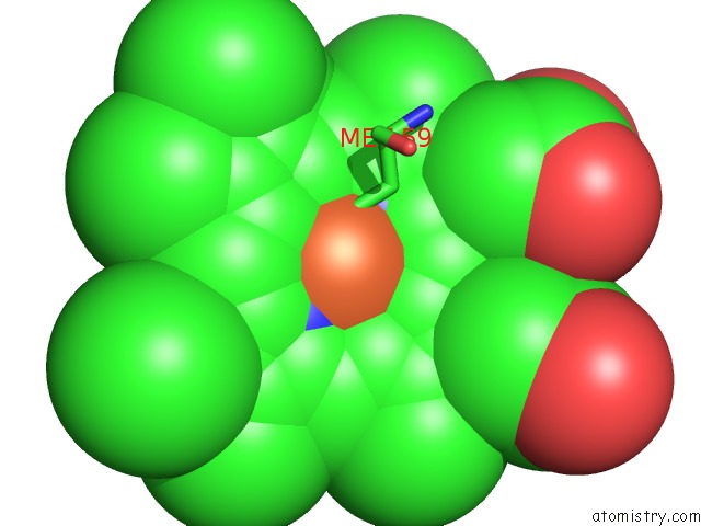
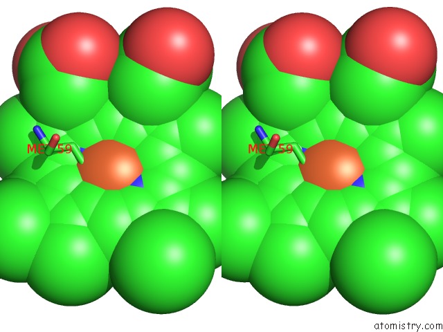
Iron binding site 1 out of 4 in 3zox
Go back to
Iron binding site 1 out
of 4 in the Crystal Structure of N64DEL Mutant of Nitrosomonas Europaea Cytochrome C552 (Monoclinic Space Group)
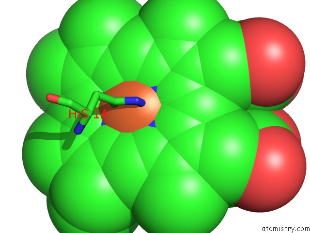
Mono view
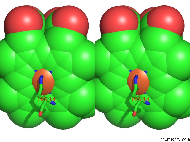
Stereo pair view
Mono view
Stereo pair view
A full contact list of Iron with other atoms in the Fe binding
site number 1 of Crystal Structure of N64DEL Mutant of Nitrosomonas Europaea Cytochrome C552 (Monoclinic Space Group) within 5.0Å range:
|
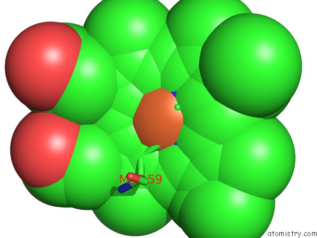
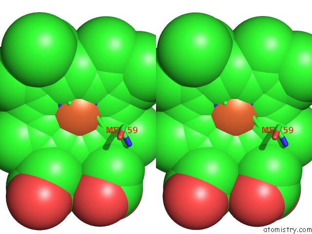
Iron binding site 2 out of 4 in 3zox
Go back to
Iron binding site 2 out
of 4 in the Crystal Structure of N64DEL Mutant of Nitrosomonas Europaea Cytochrome C552 (Monoclinic Space Group)
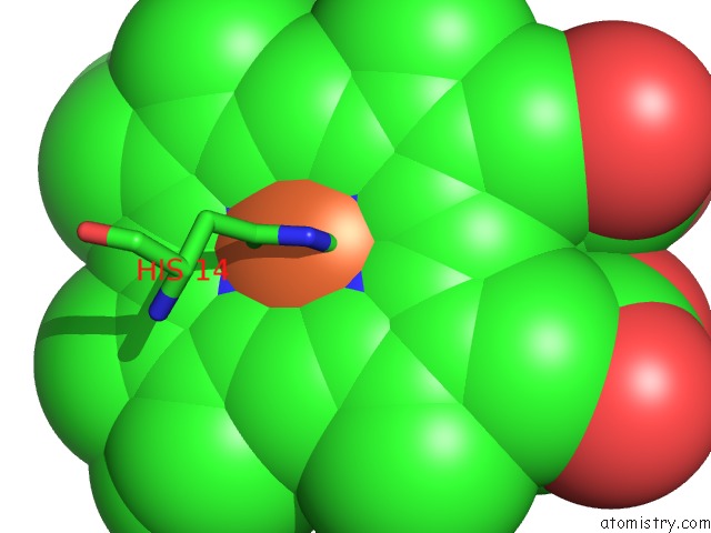
Mono view
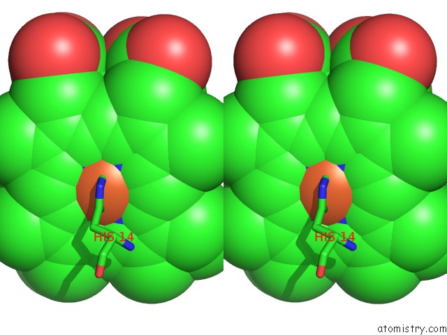
Stereo pair view
Mono view
Stereo pair view
A full contact list of Iron with other atoms in the Fe binding
site number 2 of Crystal Structure of N64DEL Mutant of Nitrosomonas Europaea Cytochrome C552 (Monoclinic Space Group) within 5.0Å range:
|
Iron binding site 3 out of 4 in 3zox
Go back to
Iron binding site 3 out
of 4 in the Crystal Structure of N64DEL Mutant of Nitrosomonas Europaea Cytochrome C552 (Monoclinic Space Group)
Mono view
Stereo pair view
Mono view
Stereo pair view
A full contact list of Iron with other atoms in the Fe binding
site number 3 of Crystal Structure of N64DEL Mutant of Nitrosomonas Europaea Cytochrome C552 (Monoclinic Space Group) within 5.0Å range:
|
Iron binding site 4 out of 4 in 3zox
Go back to
Iron binding site 4 out
of 4 in the Crystal Structure of N64DEL Mutant of Nitrosomonas Europaea Cytochrome C552 (Monoclinic Space Group)
Mono view
Stereo pair view
Mono view
Stereo pair view
A full contact list of Iron with other atoms in the Fe binding
site number 4 of Crystal Structure of N64DEL Mutant of Nitrosomonas Europaea Cytochrome C552 (Monoclinic Space Group) within 5.0Å range:
|
Reference:
M.Can,
J.Krucinska,
G.Zoppellaro,
N.H.Andersen,
J.E.Wedekind,
H.-P.Hersleth,
K.K.Andersson,
K.L.Bren.
Structural Characterization of Nitrosomonas Europaea Cytochrome C-552 Variants with Marked Differences in Electronic Structure. Chembiochem V. 14 1828 2013.
ISSN: ISSN 1439-4227
PubMed: 23908017
DOI: 10.1002/CBIC.201300118
Page generated: Sun Aug 4 23:23:44 2024
ISSN: ISSN 1439-4227
PubMed: 23908017
DOI: 10.1002/CBIC.201300118
Last articles
Cl in 5WW4Cl in 5WW3
Cl in 5WU4
Cl in 5WU3
Cl in 5WTQ
Cl in 5WU2
Cl in 5WU1
Cl in 5WTW
Cl in 5WT6
Cl in 5WT2