Iron »
PDB 3zjp-4a7m »
4a15 »
Iron in PDB 4a15: Crystal Structure of An Xpd Dna Complex
Enzymatic activity of Crystal Structure of An Xpd Dna Complex
All present enzymatic activity of Crystal Structure of An Xpd Dna Complex:
3.6.4.12;
3.6.4.12;
Protein crystallography data
The structure of Crystal Structure of An Xpd Dna Complex, PDB code: 4a15
was solved by
J.Kuper,
S.C.Wolski,
G.Michels,
C.Kisker,
with X-Ray Crystallography technique. A brief refinement statistics is given in the table below:
| Resolution Low / High (Å) | 39.53 / 2.20 |
| Space group | P 65 |
| Cell size a, b, c (Å), α, β, γ (°) | 79.060, 79.060, 175.690, 90.00, 90.00, 120.00 |
| R / Rfree (%) | 19.23 / 25.049 |
Other elements in 4a15:
The structure of Crystal Structure of An Xpd Dna Complex also contains other interesting chemical elements:
| Calcium | (Ca) | 1 atom |
Iron Binding Sites:
The binding sites of Iron atom in the Crystal Structure of An Xpd Dna Complex
(pdb code 4a15). This binding sites where shown within
5.0 Angstroms radius around Iron atom.
In total 4 binding sites of Iron where determined in the Crystal Structure of An Xpd Dna Complex, PDB code: 4a15:
Jump to Iron binding site number: 1; 2; 3; 4;
In total 4 binding sites of Iron where determined in the Crystal Structure of An Xpd Dna Complex, PDB code: 4a15:
Jump to Iron binding site number: 1; 2; 3; 4;
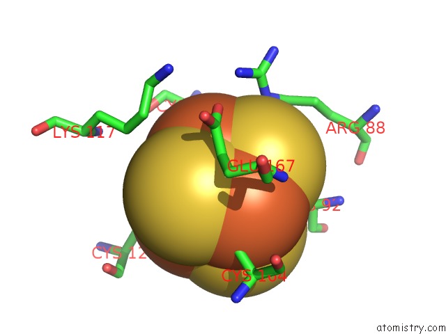
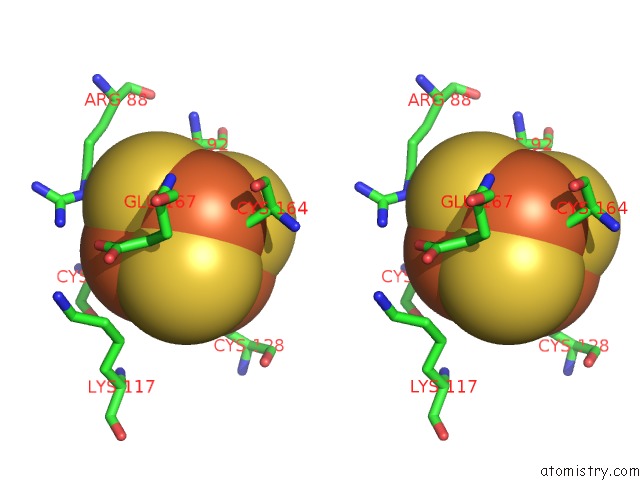
Iron binding site 1 out of 4 in 4a15
Go back to
Iron binding site 1 out
of 4 in the Crystal Structure of An Xpd Dna Complex
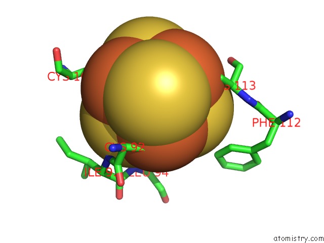
Mono view
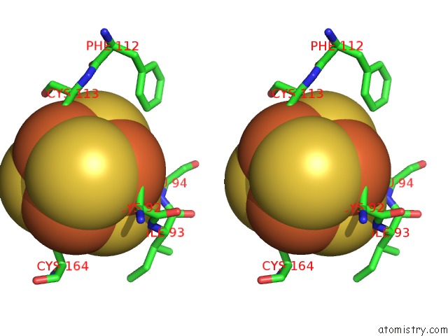
Stereo pair view
Mono view
Stereo pair view
A full contact list of Iron with other atoms in the Fe binding
site number 1 of Crystal Structure of An Xpd Dna Complex within 5.0Å range:
|
Iron binding site 2 out of 4 in 4a15
Go back to
Iron binding site 2 out
of 4 in the Crystal Structure of An Xpd Dna Complex
Mono view
Stereo pair view
Mono view
Stereo pair view
A full contact list of Iron with other atoms in the Fe binding
site number 2 of Crystal Structure of An Xpd Dna Complex within 5.0Å range:
|
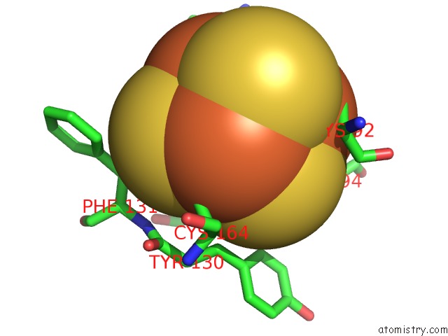
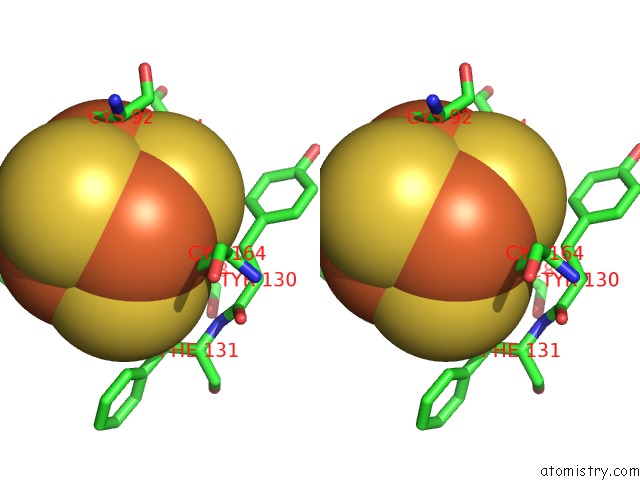
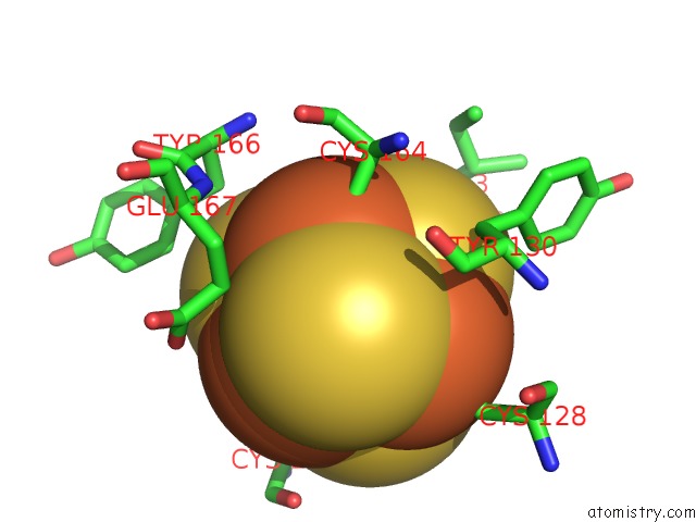
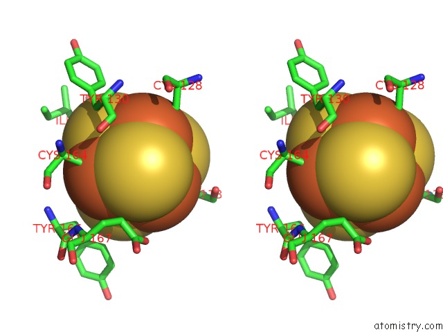
Iron binding site 3 out of 4 in 4a15
Go back to
Iron binding site 3 out
of 4 in the Crystal Structure of An Xpd Dna Complex
Mono view
Stereo pair view
Mono view
Stereo pair view
A full contact list of Iron with other atoms in the Fe binding
site number 3 of Crystal Structure of An Xpd Dna Complex within 5.0Å range:
|
Iron binding site 4 out of 4 in 4a15
Go back to
Iron binding site 4 out
of 4 in the Crystal Structure of An Xpd Dna Complex
Mono view
Stereo pair view
Mono view
Stereo pair view
A full contact list of Iron with other atoms in the Fe binding
site number 4 of Crystal Structure of An Xpd Dna Complex within 5.0Å range:
|
Reference:
J.Kuper,
S.C.Wolski,
G.Michels,
C.Kisker.
Functional and Structural Studies of the Nucleotide Excision Repair Helicase Xpd Suggest A Polarity For Dna Translocation. Embo J. V. 31 494 2011.
ISSN: ISSN 0261-4189
PubMed: 22081108
DOI: 10.1038/EMBOJ.2011.374
Page generated: Sun Aug 4 23:28:35 2024
ISSN: ISSN 0261-4189
PubMed: 22081108
DOI: 10.1038/EMBOJ.2011.374
Last articles
Zn in 9MJ5Zn in 9HNW
Zn in 9G0L
Zn in 9FNE
Zn in 9DZN
Zn in 9E0I
Zn in 9D32
Zn in 9DAK
Zn in 8ZXC
Zn in 8ZUF