Iron »
PDB 3zjp-4a7m »
4a6z »
Iron in PDB 4a6z: Cytochrome C Peroxidase with Bound Guaiacol
Enzymatic activity of Cytochrome C Peroxidase with Bound Guaiacol
All present enzymatic activity of Cytochrome C Peroxidase with Bound Guaiacol:
1.11.1.5;
1.11.1.5;
Protein crystallography data
The structure of Cytochrome C Peroxidase with Bound Guaiacol, PDB code: 4a6z
was solved by
E.J.Murphy,
C.L.Metcalfe,
C.Nnamchi,
E.L.Raven,
P.C.E.Moody,
with X-Ray Crystallography technique. A brief refinement statistics is given in the table below:
| Resolution Low / High (Å) | 27.24 / 1.61 |
| Space group | P 21 21 21 |
| Cell size a, b, c (Å), α, β, γ (°) | 51.106, 74.990, 106.903, 90.00, 90.00, 90.00 |
| R / Rfree (%) | 17.7 / 21 |
Iron Binding Sites:
The binding sites of Iron atom in the Cytochrome C Peroxidase with Bound Guaiacol
(pdb code 4a6z). This binding sites where shown within
5.0 Angstroms radius around Iron atom.
In total only one binding site of Iron was determined in the Cytochrome C Peroxidase with Bound Guaiacol, PDB code: 4a6z:
In total only one binding site of Iron was determined in the Cytochrome C Peroxidase with Bound Guaiacol, PDB code: 4a6z:
Iron binding site 1 out of 1 in 4a6z
Go back to
Iron binding site 1 out
of 1 in the Cytochrome C Peroxidase with Bound Guaiacol
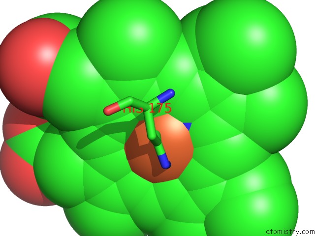
Mono view
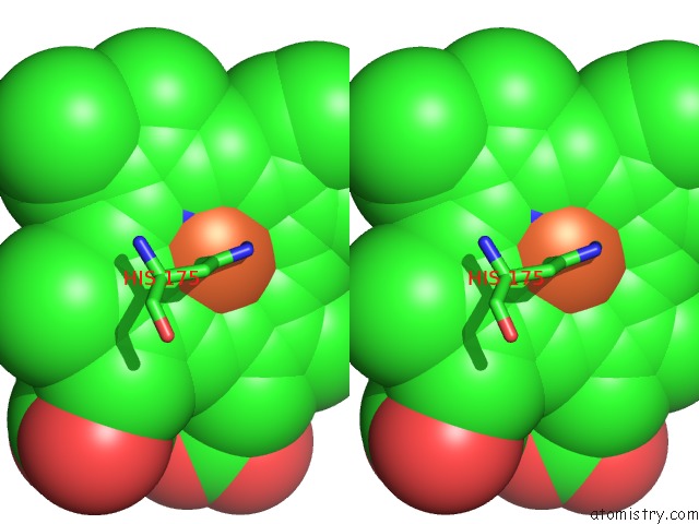
Stereo pair view
Mono view
Stereo pair view
A full contact list of Iron with other atoms in the Fe binding
site number 1 of Cytochrome C Peroxidase with Bound Guaiacol within 5.0Å range:
|
Reference:
E.J.Murphy,
C.L.Metcalfe,
C.Nnamchi,
P.C.E.Moody,
E.L.Raven.
Crystal Structure of Guaiacol and Phenol Bound to A Heme Peroxidase. Febs J. V. 279 1632 2012.
ISSN: ISSN 1742-464X
PubMed: 22093282
DOI: 10.1111/J.1742-4658.2011.08425.X
Page generated: Sun Aug 4 23:28:46 2024
ISSN: ISSN 1742-464X
PubMed: 22093282
DOI: 10.1111/J.1742-4658.2011.08425.X
Last articles
Zn in 9JYWZn in 9IR4
Zn in 9IR3
Zn in 9GMX
Zn in 9GMW
Zn in 9JEJ
Zn in 9ERF
Zn in 9ERE
Zn in 9EGV
Zn in 9EGW