Iron »
PDB 4ccx-4cun »
4cdp »
Iron in PDB 4cdp: Improved Coordinates For Escherichia Coli O157:H7 Heme Degrading Enzyme Chus.
Protein crystallography data
The structure of Improved Coordinates For Escherichia Coli O157:H7 Heme Degrading Enzyme Chus., PDB code: 4cdp
was solved by
M.D.L.Suits,
N.Jaffer,
Z.Jia,
with X-Ray Crystallography technique. A brief refinement statistics is given in the table below:
| Resolution Low / High (Å) | 30.08 / 1.45 |
| Space group | H 3 |
| Cell size a, b, c (Å), α, β, γ (°) | 106.487, 106.487, 90.229, 90.00, 90.00, 120.00 |
| R / Rfree (%) | 13.259 / 17.253 |
Iron Binding Sites:
The binding sites of Iron atom in the Improved Coordinates For Escherichia Coli O157:H7 Heme Degrading Enzyme Chus.
(pdb code 4cdp). This binding sites where shown within
5.0 Angstroms radius around Iron atom.
In total only one binding site of Iron was determined in the Improved Coordinates For Escherichia Coli O157:H7 Heme Degrading Enzyme Chus., PDB code: 4cdp:
In total only one binding site of Iron was determined in the Improved Coordinates For Escherichia Coli O157:H7 Heme Degrading Enzyme Chus., PDB code: 4cdp:
Iron binding site 1 out of 1 in 4cdp
Go back to
Iron binding site 1 out
of 1 in the Improved Coordinates For Escherichia Coli O157:H7 Heme Degrading Enzyme Chus.
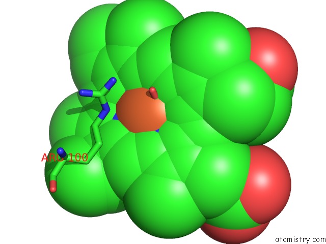
Mono view
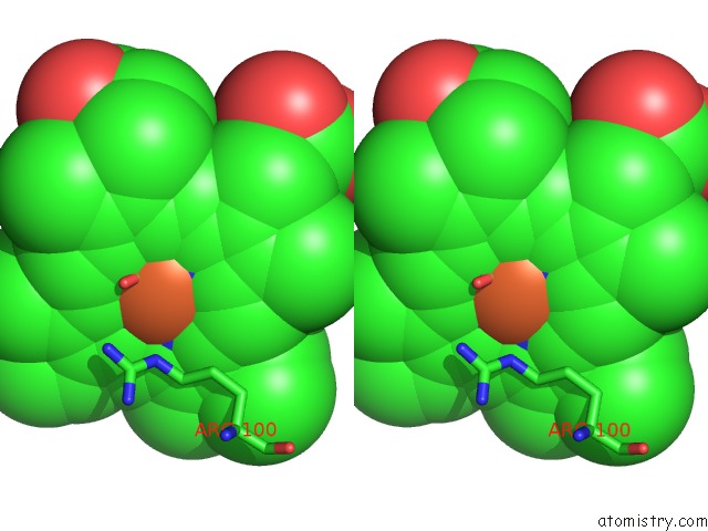
Stereo pair view
Mono view
Stereo pair view
A full contact list of Iron with other atoms in the Fe binding
site number 1 of Improved Coordinates For Escherichia Coli O157:H7 Heme Degrading Enzyme Chus. within 5.0Å range:
|
Reference:
M.D.Suits,
N.Jaffer,
Z.Jia.
Structure of the Escherichia Coli O157:H7 Heme Oxygenase Chus in Complex with Heme and Enzymatic Inactivation By Mutation of the Heme Coordinating Residue His-193. J.Biol.Chem. V. 281 36776 2006.
ISSN: ISSN 0021-9258
PubMed: 17023414
DOI: 10.1074/JBC.M607684200
Page generated: Mon Aug 5 00:34:59 2024
ISSN: ISSN 0021-9258
PubMed: 17023414
DOI: 10.1074/JBC.M607684200
Last articles
Zn in 9MJ5Zn in 9HNW
Zn in 9G0L
Zn in 9FNE
Zn in 9DZN
Zn in 9E0I
Zn in 9D32
Zn in 9DAK
Zn in 8ZXC
Zn in 8ZUF