Iron »
PDB 4ccx-4cun »
4ceh »
Iron in PDB 4ceh: Crystal Structure of Addab with A Forked Dna Substrate
Enzymatic activity of Crystal Structure of Addab with A Forked Dna Substrate
All present enzymatic activity of Crystal Structure of Addab with A Forked Dna Substrate:
3.6.4.12;
3.6.4.12;
Protein crystallography data
The structure of Crystal Structure of Addab with A Forked Dna Substrate, PDB code: 4ceh
was solved by
W.W.Krajewski,
M.Wilkinson,
X.Fu,
N.B.Cronin,
D.Wigley,
with X-Ray Crystallography technique. A brief refinement statistics is given in the table below:
| Resolution Low / High (Å) | 29.939 / 3.24 |
| Space group | P 1 |
| Cell size a, b, c (Å), α, β, γ (°) | 77.373, 96.766, 109.693, 104.38, 96.11, 90.03 |
| R / Rfree (%) | 22.92 / 26.38 |
Iron Binding Sites:
The binding sites of Iron atom in the Crystal Structure of Addab with A Forked Dna Substrate
(pdb code 4ceh). This binding sites where shown within
5.0 Angstroms radius around Iron atom.
In total 4 binding sites of Iron where determined in the Crystal Structure of Addab with A Forked Dna Substrate, PDB code: 4ceh:
Jump to Iron binding site number: 1; 2; 3; 4;
In total 4 binding sites of Iron where determined in the Crystal Structure of Addab with A Forked Dna Substrate, PDB code: 4ceh:
Jump to Iron binding site number: 1; 2; 3; 4;
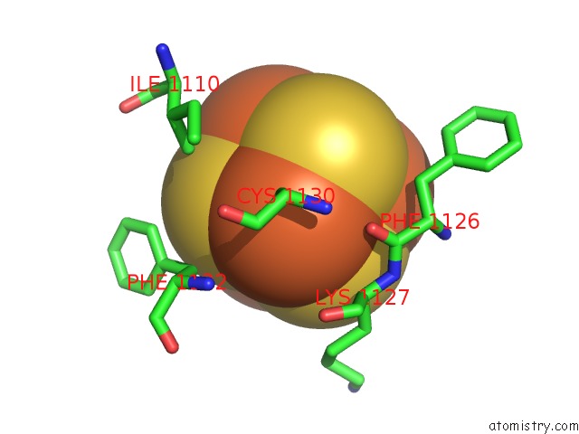
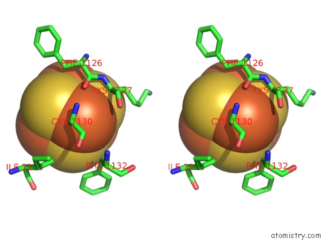
Iron binding site 1 out of 4 in 4ceh
Go back to
Iron binding site 1 out
of 4 in the Crystal Structure of Addab with A Forked Dna Substrate
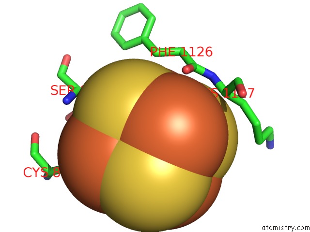
Mono view
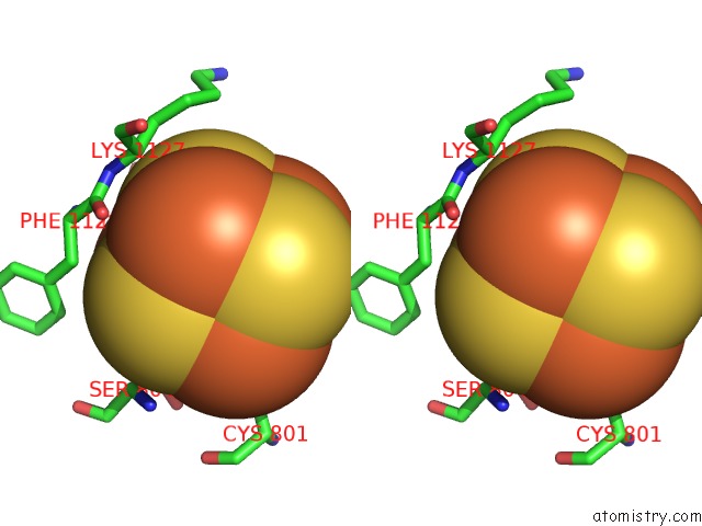
Stereo pair view
Mono view
Stereo pair view
A full contact list of Iron with other atoms in the Fe binding
site number 1 of Crystal Structure of Addab with A Forked Dna Substrate within 5.0Å range:
|
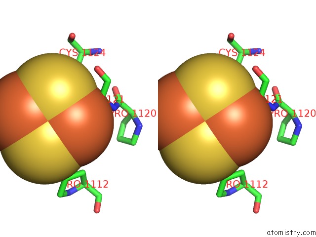
Iron binding site 2 out of 4 in 4ceh
Go back to
Iron binding site 2 out
of 4 in the Crystal Structure of Addab with A Forked Dna Substrate
Mono view
Stereo pair view
Mono view
Stereo pair view
A full contact list of Iron with other atoms in the Fe binding
site number 2 of Crystal Structure of Addab with A Forked Dna Substrate within 5.0Å range:
|
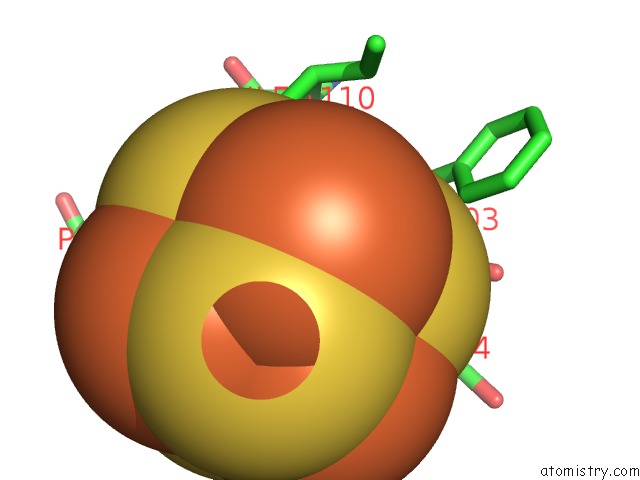
Iron binding site 3 out of 4 in 4ceh
Go back to
Iron binding site 3 out
of 4 in the Crystal Structure of Addab with A Forked Dna Substrate
Mono view
Stereo pair view
Mono view
Stereo pair view
A full contact list of Iron with other atoms in the Fe binding
site number 3 of Crystal Structure of Addab with A Forked Dna Substrate within 5.0Å range:
|
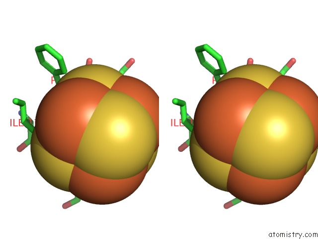
Iron binding site 4 out of 4 in 4ceh
Go back to
Iron binding site 4 out
of 4 in the Crystal Structure of Addab with A Forked Dna Substrate

Mono view
Stereo pair view
Mono view
Stereo pair view
A full contact list of Iron with other atoms in the Fe binding
site number 4 of Crystal Structure of Addab with A Forked Dna Substrate within 5.0Å range:
|
Reference:
W.W.Krajewski,
X.Fu,
M.Wilkinson,
N.B.Cronin,
M.S.Dillingham,
D.B.Wigley.
Structural Basis For Translocation By Addab Helicase-Nuclease and Its Arrest at Chi Sites. Nature V. 508 416 2014.
ISSN: ISSN 0028-0836
PubMed: 24670664
DOI: 10.1038/NATURE13037
Page generated: Mon Aug 5 00:35:00 2024
ISSN: ISSN 0028-0836
PubMed: 24670664
DOI: 10.1038/NATURE13037
Last articles
Zn in 9J0NZn in 9J0O
Zn in 9J0P
Zn in 9FJX
Zn in 9EKB
Zn in 9C0F
Zn in 9CAH
Zn in 9CH0
Zn in 9CH3
Zn in 9CH1