Iron »
PDB 4d37-4di0 »
4d7d »
Iron in PDB 4d7d: Cytochrome P450 3A4 Bound to An Inhibitor
Enzymatic activity of Cytochrome P450 3A4 Bound to An Inhibitor
All present enzymatic activity of Cytochrome P450 3A4 Bound to An Inhibitor:
1.14.13.157; 1.14.13.32; 1.14.13.67; 1.14.13.97;
1.14.13.157; 1.14.13.32; 1.14.13.67; 1.14.13.97;
Protein crystallography data
The structure of Cytochrome P450 3A4 Bound to An Inhibitor, PDB code: 4d7d
was solved by
I.Sevrioukova,
T.Poulos,
with X-Ray Crystallography technique. A brief refinement statistics is given in the table below:
| Resolution Low / High (Å) | 37.44 / 2.76 |
| Space group | I 2 2 2 |
| Cell size a, b, c (Å), α, β, γ (°) | 78.138, 101.332, 127.966, 90.00, 90.00, 90.00 |
| R / Rfree (%) | 20 / 27.8 |
Iron Binding Sites:
The binding sites of Iron atom in the Cytochrome P450 3A4 Bound to An Inhibitor
(pdb code 4d7d). This binding sites where shown within
5.0 Angstroms radius around Iron atom.
In total only one binding site of Iron was determined in the Cytochrome P450 3A4 Bound to An Inhibitor, PDB code: 4d7d:
In total only one binding site of Iron was determined in the Cytochrome P450 3A4 Bound to An Inhibitor, PDB code: 4d7d:
Iron binding site 1 out of 1 in 4d7d
Go back to
Iron binding site 1 out
of 1 in the Cytochrome P450 3A4 Bound to An Inhibitor
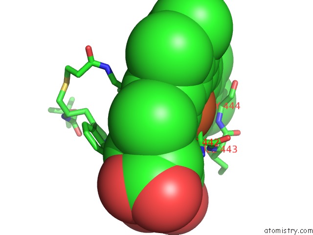
Mono view
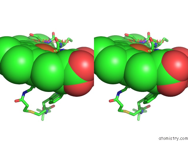
Stereo pair view
Mono view
Stereo pair view
A full contact list of Iron with other atoms in the Fe binding
site number 1 of Cytochrome P450 3A4 Bound to An Inhibitor within 5.0Å range:
|
Reference:
P.Kaur,
R.Chamberlin,
T.L.Poulos,
I.F.Sevrioukova.
Structure-Based Inhibitor Design For Evaluation of A CYP3A4 Pharmacophore Model. J.Med.Chem. V. 59 4210 2016.
ISSN: ISSN 0022-2623
PubMed: 26371436
DOI: 10.1021/ACS.JMEDCHEM.5B01146
Page generated: Mon Aug 5 01:11:30 2024
ISSN: ISSN 0022-2623
PubMed: 26371436
DOI: 10.1021/ACS.JMEDCHEM.5B01146
Last articles
Zn in 9J0NZn in 9J0O
Zn in 9J0P
Zn in 9FJX
Zn in 9EKB
Zn in 9C0F
Zn in 9CAH
Zn in 9CH0
Zn in 9CH3
Zn in 9CH1