Iron »
PDB 4d37-4di0 »
4dc8 »
Iron in PDB 4dc8: Crystal Structure of Myoglobin Unexposed to Excessive Sonicc Imaging Laser Dose.
Protein crystallography data
The structure of Crystal Structure of Myoglobin Unexposed to Excessive Sonicc Imaging Laser Dose., PDB code: 4dc8
was solved by
M.Becker,
A.M.Mulichak,
D.J.Kissick,
R.F.Fischetti,
L.J.Keefe,
D.J.Simpson,
with X-Ray Crystallography technique. A brief refinement statistics is given in the table below:
| Resolution Low / High (Å) | 33.99 / 1.50 |
| Space group | P 1 21 1 |
| Cell size a, b, c (Å), α, β, γ (°) | 35.363, 28.794, 63.056, 90.00, 106.05, 90.00 |
| R / Rfree (%) | 18.3 / 21.8 |
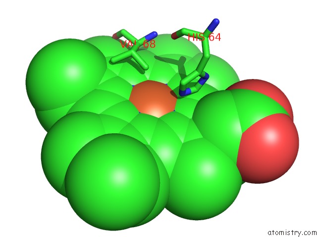
Iron Binding Sites:
The binding sites of Iron atom in the Crystal Structure of Myoglobin Unexposed to Excessive Sonicc Imaging Laser Dose.
(pdb code 4dc8). This binding sites where shown within
5.0 Angstroms radius around Iron atom.
In total only one binding site of Iron was determined in the Crystal Structure of Myoglobin Unexposed to Excessive Sonicc Imaging Laser Dose., PDB code: 4dc8:
In total only one binding site of Iron was determined in the Crystal Structure of Myoglobin Unexposed to Excessive Sonicc Imaging Laser Dose., PDB code: 4dc8:
Iron binding site 1 out of 1 in 4dc8
Go back to
Iron binding site 1 out
of 1 in the Crystal Structure of Myoglobin Unexposed to Excessive Sonicc Imaging Laser Dose.
Mono view

Stereo pair view
Mono view
Stereo pair view
A full contact list of Iron with other atoms in the Fe binding
site number 1 of Crystal Structure of Myoglobin Unexposed to Excessive Sonicc Imaging Laser Dose. within 5.0Å range:
|
Reference:
D.J.Kissick,
C.M.Dettmar,
M.Becker,
A.M.Mulichak,
V.Cherezov,
S.L.Ginell,
K.P.Battaile,
L.J.Keefe,
R.F.Fischetti,
G.J.Simpson.
Towards Protein-Crystal Centering Using Second-Harmonic Generation (Shg) Microscopy. Acta Crystallogr.,Sect.D V. 69 843 2013.
ISSN: ISSN 0907-4449
PubMed: 23633594
DOI: 10.1107/S0907444913002746
Page generated: Mon Aug 5 01:13:31 2024
ISSN: ISSN 0907-4449
PubMed: 23633594
DOI: 10.1107/S0907444913002746
Last articles
Zn in 9J0NZn in 9J0O
Zn in 9J0P
Zn in 9FJX
Zn in 9EKB
Zn in 9C0F
Zn in 9CAH
Zn in 9CH0
Zn in 9CH3
Zn in 9CH1