Iron »
PDB 4dig-4egm »
4djf »
Iron in PDB 4djf: Crystal Structure of Folate-Bound Corrinoid Iron-Sulfur Protein (Cfesp) in Complex with Its Methyltransferase (Metr), Co-Crystallized with Folate and Ti(III) Citrate Reductant
Protein crystallography data
The structure of Crystal Structure of Folate-Bound Corrinoid Iron-Sulfur Protein (Cfesp) in Complex with Its Methyltransferase (Metr), Co-Crystallized with Folate and Ti(III) Citrate Reductant, PDB code: 4djf
was solved by
Y.Kung,
C.L.Drennan,
with X-Ray Crystallography technique. A brief refinement statistics is given in the table below:
| Resolution Low / High (Å) | 49.75 / 3.03 |
| Space group | P 21 21 2 |
| Cell size a, b, c (Å), α, β, γ (°) | 136.006, 250.686, 81.800, 90.00, 90.00, 90.00 |
| R / Rfree (%) | 26.9 / 30.8 |
Other elements in 4djf:
The structure of Crystal Structure of Folate-Bound Corrinoid Iron-Sulfur Protein (Cfesp) in Complex with Its Methyltransferase (Metr), Co-Crystallized with Folate and Ti(III) Citrate Reductant also contains other interesting chemical elements:
| Cobalt | (Co) | 2 atoms |
| Calcium | (Ca) | 2 atoms |
Iron Binding Sites:
The binding sites of Iron atom in the Crystal Structure of Folate-Bound Corrinoid Iron-Sulfur Protein (Cfesp) in Complex with Its Methyltransferase (Metr), Co-Crystallized with Folate and Ti(III) Citrate Reductant
(pdb code 4djf). This binding sites where shown within
5.0 Angstroms radius around Iron atom.
In total 8 binding sites of Iron where determined in the Crystal Structure of Folate-Bound Corrinoid Iron-Sulfur Protein (Cfesp) in Complex with Its Methyltransferase (Metr), Co-Crystallized with Folate and Ti(III) Citrate Reductant, PDB code: 4djf:
Jump to Iron binding site number: 1; 2; 3; 4; 5; 6; 7; 8;
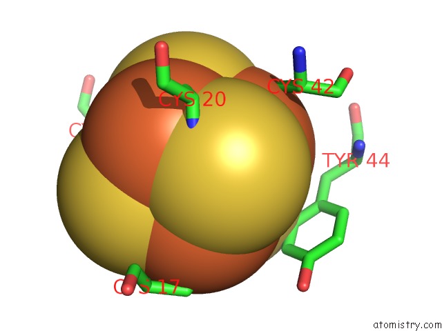
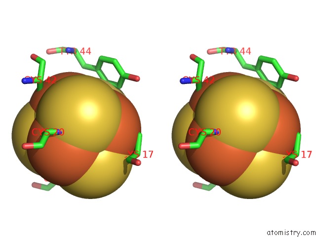
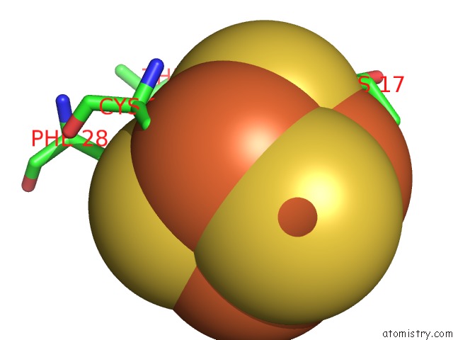
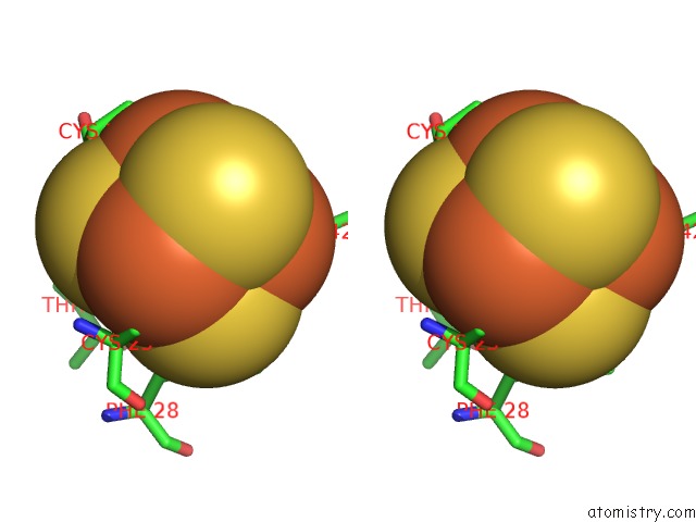
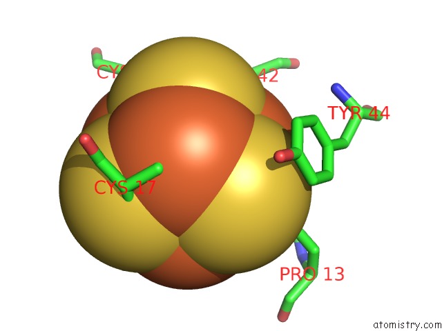
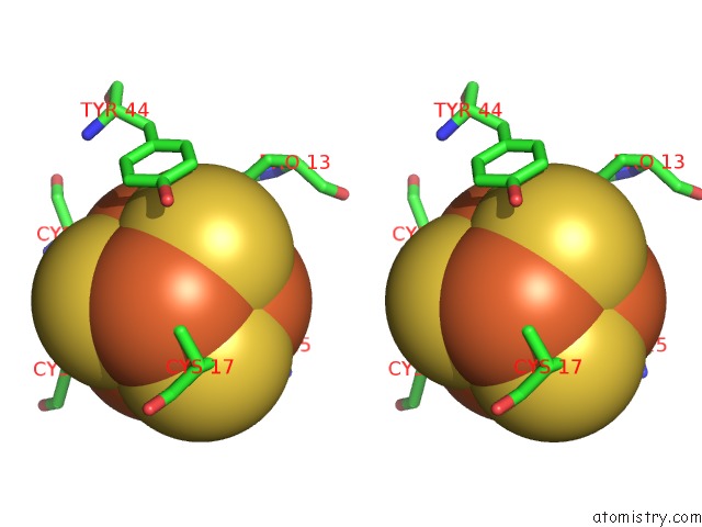
Iron binding site 1 out of 8 in 4djf
Go back to
Iron binding site 1 out
of 8 in the Crystal Structure of Folate-Bound Corrinoid Iron-Sulfur Protein (Cfesp) in Complex with Its Methyltransferase (Metr), Co-Crystallized with Folate and Ti(III) Citrate Reductant
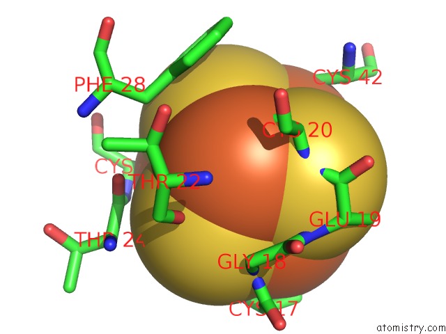
Mono view
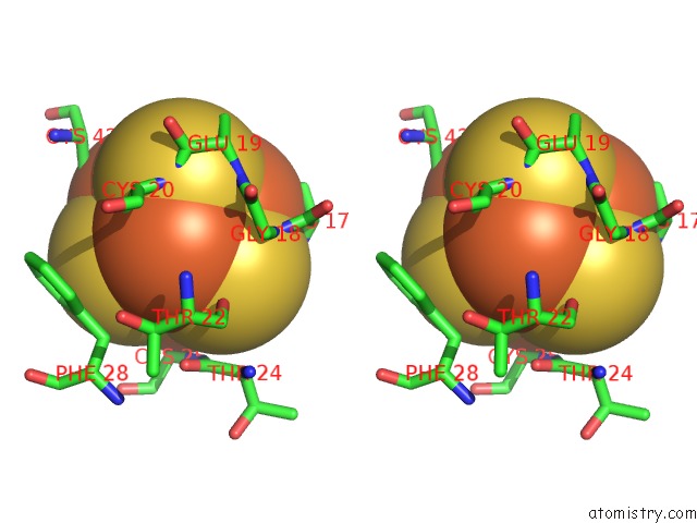
Stereo pair view
Mono view
Stereo pair view
A full contact list of Iron with other atoms in the Fe binding
site number 1 of Crystal Structure of Folate-Bound Corrinoid Iron-Sulfur Protein (Cfesp) in Complex with Its Methyltransferase (Metr), Co-Crystallized with Folate and Ti(III) Citrate Reductant within 5.0Å range:
|
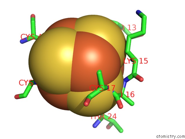
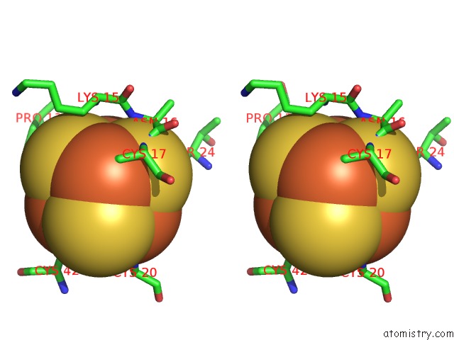
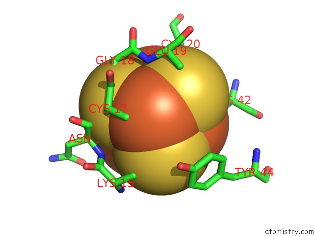
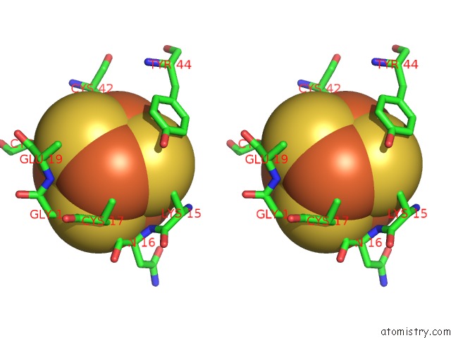
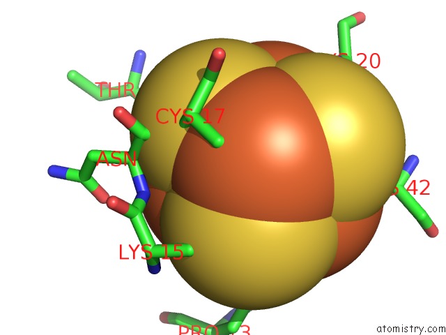
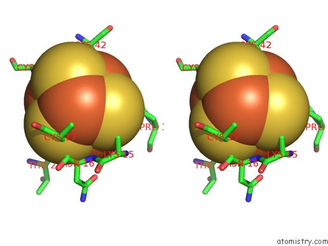
Iron binding site 2 out of 8 in 4djf
Go back to
Iron binding site 2 out
of 8 in the Crystal Structure of Folate-Bound Corrinoid Iron-Sulfur Protein (Cfesp) in Complex with Its Methyltransferase (Metr), Co-Crystallized with Folate and Ti(III) Citrate Reductant
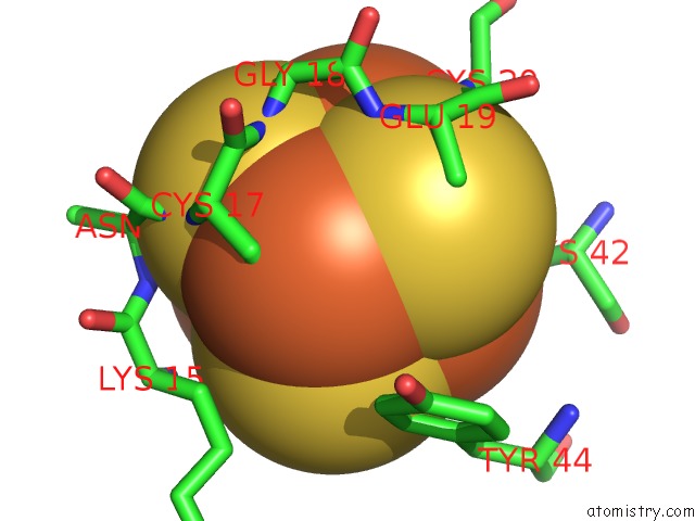
Mono view
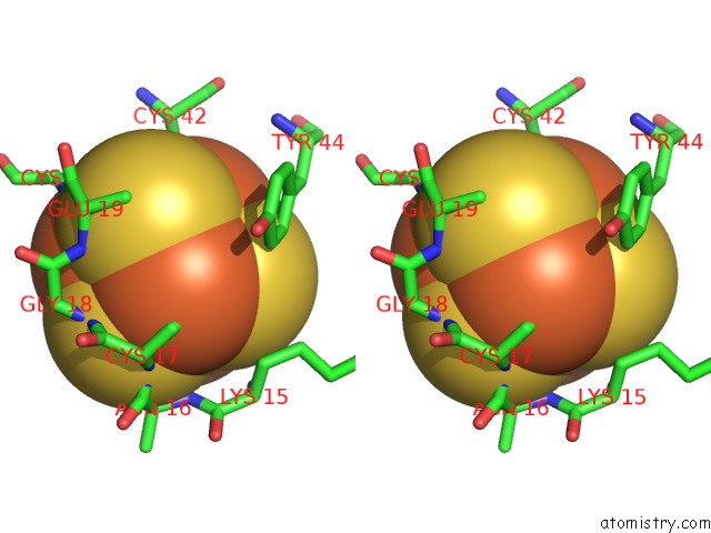
Stereo pair view
Mono view
Stereo pair view
A full contact list of Iron with other atoms in the Fe binding
site number 2 of Crystal Structure of Folate-Bound Corrinoid Iron-Sulfur Protein (Cfesp) in Complex with Its Methyltransferase (Metr), Co-Crystallized with Folate and Ti(III) Citrate Reductant within 5.0Å range:
|
Iron binding site 3 out of 8 in 4djf
Go back to
Iron binding site 3 out
of 8 in the Crystal Structure of Folate-Bound Corrinoid Iron-Sulfur Protein (Cfesp) in Complex with Its Methyltransferase (Metr), Co-Crystallized with Folate and Ti(III) Citrate Reductant
Mono view
Stereo pair view
Mono view
Stereo pair view
A full contact list of Iron with other atoms in the Fe binding
site number 3 of Crystal Structure of Folate-Bound Corrinoid Iron-Sulfur Protein (Cfesp) in Complex with Its Methyltransferase (Metr), Co-Crystallized with Folate and Ti(III) Citrate Reductant within 5.0Å range:
|
Iron binding site 4 out of 8 in 4djf
Go back to
Iron binding site 4 out
of 8 in the Crystal Structure of Folate-Bound Corrinoid Iron-Sulfur Protein (Cfesp) in Complex with Its Methyltransferase (Metr), Co-Crystallized with Folate and Ti(III) Citrate Reductant
Mono view
Stereo pair view
Mono view
Stereo pair view
A full contact list of Iron with other atoms in the Fe binding
site number 4 of Crystal Structure of Folate-Bound Corrinoid Iron-Sulfur Protein (Cfesp) in Complex with Its Methyltransferase (Metr), Co-Crystallized with Folate and Ti(III) Citrate Reductant within 5.0Å range:
|
Iron binding site 5 out of 8 in 4djf
Go back to
Iron binding site 5 out
of 8 in the Crystal Structure of Folate-Bound Corrinoid Iron-Sulfur Protein (Cfesp) in Complex with Its Methyltransferase (Metr), Co-Crystallized with Folate and Ti(III) Citrate Reductant
Mono view
Stereo pair view
Mono view
Stereo pair view
A full contact list of Iron with other atoms in the Fe binding
site number 5 of Crystal Structure of Folate-Bound Corrinoid Iron-Sulfur Protein (Cfesp) in Complex with Its Methyltransferase (Metr), Co-Crystallized with Folate and Ti(III) Citrate Reductant within 5.0Å range:
|
Iron binding site 6 out of 8 in 4djf
Go back to
Iron binding site 6 out
of 8 in the Crystal Structure of Folate-Bound Corrinoid Iron-Sulfur Protein (Cfesp) in Complex with Its Methyltransferase (Metr), Co-Crystallized with Folate and Ti(III) Citrate Reductant
Mono view
Stereo pair view
Mono view
Stereo pair view
A full contact list of Iron with other atoms in the Fe binding
site number 6 of Crystal Structure of Folate-Bound Corrinoid Iron-Sulfur Protein (Cfesp) in Complex with Its Methyltransferase (Metr), Co-Crystallized with Folate and Ti(III) Citrate Reductant within 5.0Å range:
|
Iron binding site 7 out of 8 in 4djf
Go back to
Iron binding site 7 out
of 8 in the Crystal Structure of Folate-Bound Corrinoid Iron-Sulfur Protein (Cfesp) in Complex with Its Methyltransferase (Metr), Co-Crystallized with Folate and Ti(III) Citrate Reductant
Mono view
Stereo pair view
Mono view
Stereo pair view
A full contact list of Iron with other atoms in the Fe binding
site number 7 of Crystal Structure of Folate-Bound Corrinoid Iron-Sulfur Protein (Cfesp) in Complex with Its Methyltransferase (Metr), Co-Crystallized with Folate and Ti(III) Citrate Reductant within 5.0Å range:
|
Iron binding site 8 out of 8 in 4djf
Go back to
Iron binding site 8 out
of 8 in the Crystal Structure of Folate-Bound Corrinoid Iron-Sulfur Protein (Cfesp) in Complex with Its Methyltransferase (Metr), Co-Crystallized with Folate and Ti(III) Citrate Reductant
Mono view
Stereo pair view
Mono view
Stereo pair view
A full contact list of Iron with other atoms in the Fe binding
site number 8 of Crystal Structure of Folate-Bound Corrinoid Iron-Sulfur Protein (Cfesp) in Complex with Its Methyltransferase (Metr), Co-Crystallized with Folate and Ti(III) Citrate Reductant within 5.0Å range:
|
Reference:
Y.Kung,
N.Ando,
T.I.Doukov,
L.C.Blasiak,
G.Bender,
J.Seravalli,
S.W.Ragsdale,
C.L.Drennan.
Visualizing Molecular Juggling Within A B12-Dependent Methyltransferase Complex. Nature V. 484 265 2012.
ISSN: ISSN 0028-0836
PubMed: 22419154
DOI: 10.1038/NATURE10916
Page generated: Mon Aug 5 01:18:47 2024
ISSN: ISSN 0028-0836
PubMed: 22419154
DOI: 10.1038/NATURE10916
Last articles
Fe in 2YXOFe in 2YRS
Fe in 2YXC
Fe in 2YNM
Fe in 2YVJ
Fe in 2YP1
Fe in 2YU2
Fe in 2YU1
Fe in 2YQB
Fe in 2YOO