Iron »
PDB 4dig-4egm »
4dl1 »
Iron in PDB 4dl1: Crystal Structure of Human Myeloperoxidase with Covalent Thioxanthine Analog
Enzymatic activity of Crystal Structure of Human Myeloperoxidase with Covalent Thioxanthine Analog
All present enzymatic activity of Crystal Structure of Human Myeloperoxidase with Covalent Thioxanthine Analog:
1.11.2.2;
1.11.2.2;
Protein crystallography data
The structure of Crystal Structure of Human Myeloperoxidase with Covalent Thioxanthine Analog, PDB code: 4dl1
was solved by
F.Vajdos,
A.Varghese,
with X-Ray Crystallography technique. A brief refinement statistics is given in the table below:
| Resolution Low / High (Å) | 128.49 / 2.00 |
| Space group | P 1 21 1 |
| Cell size a, b, c (Å), α, β, γ (°) | 63.829, 242.636, 151.505, 90.00, 91.19, 90.00 |
| R / Rfree (%) | 19 / 24.6 |
Other elements in 4dl1:
The structure of Crystal Structure of Human Myeloperoxidase with Covalent Thioxanthine Analog also contains other interesting chemical elements:
| Chlorine | (Cl) | 8 atoms |
| Calcium | (Ca) | 8 atoms |
Iron Binding Sites:
The binding sites of Iron atom in the Crystal Structure of Human Myeloperoxidase with Covalent Thioxanthine Analog
(pdb code 4dl1). This binding sites where shown within
5.0 Angstroms radius around Iron atom.
In total 8 binding sites of Iron where determined in the Crystal Structure of Human Myeloperoxidase with Covalent Thioxanthine Analog, PDB code: 4dl1:
Jump to Iron binding site number: 1; 2; 3; 4; 5; 6; 7; 8;
In total 8 binding sites of Iron where determined in the Crystal Structure of Human Myeloperoxidase with Covalent Thioxanthine Analog, PDB code: 4dl1:
Jump to Iron binding site number: 1; 2; 3; 4; 5; 6; 7; 8;
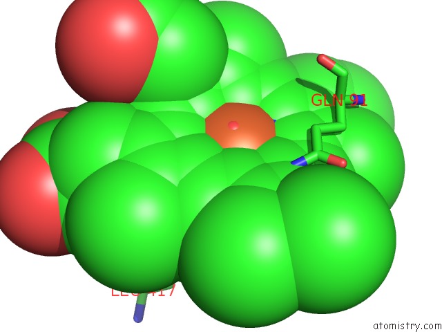
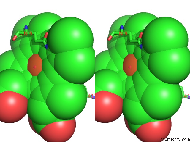
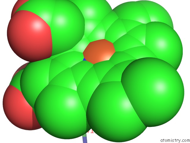
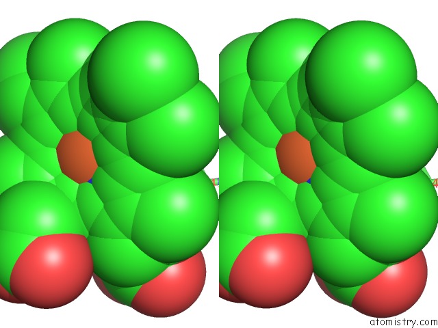
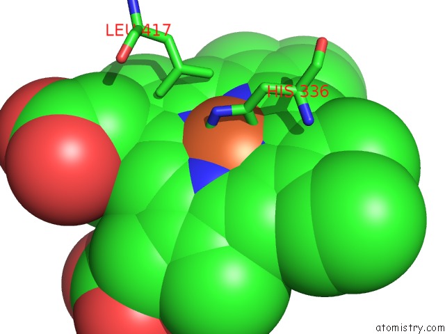
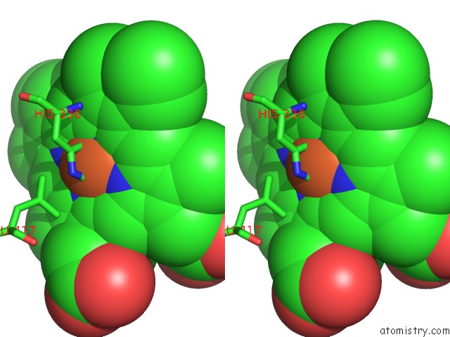
Iron binding site 1 out of 8 in 4dl1
Go back to
Iron binding site 1 out
of 8 in the Crystal Structure of Human Myeloperoxidase with Covalent Thioxanthine Analog
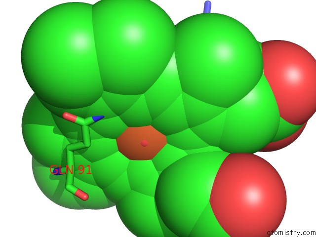
Mono view
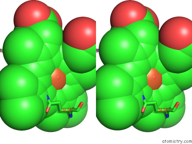
Stereo pair view
Mono view
Stereo pair view
A full contact list of Iron with other atoms in the Fe binding
site number 1 of Crystal Structure of Human Myeloperoxidase with Covalent Thioxanthine Analog within 5.0Å range:
|
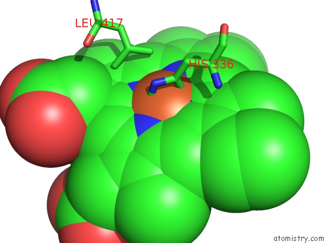
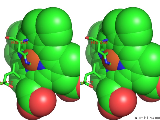
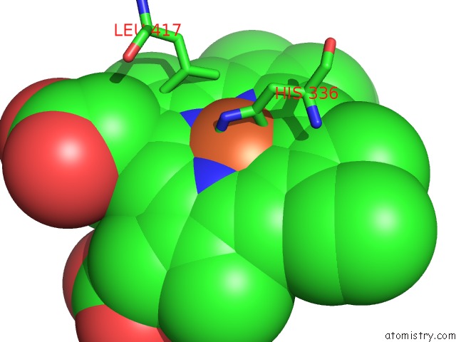
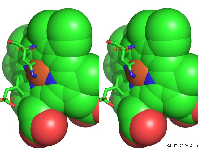
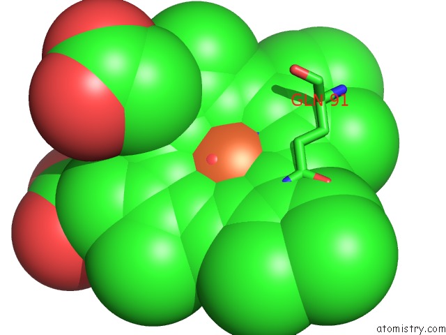
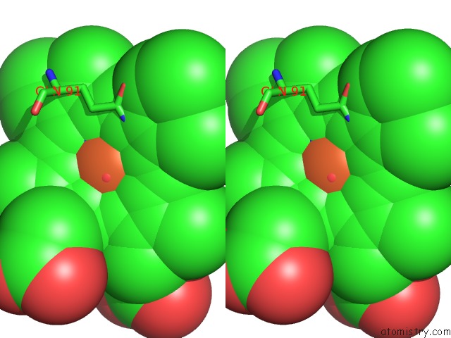
Iron binding site 2 out of 8 in 4dl1
Go back to
Iron binding site 2 out
of 8 in the Crystal Structure of Human Myeloperoxidase with Covalent Thioxanthine Analog
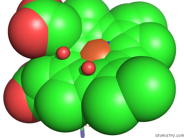
Mono view
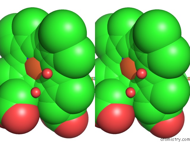
Stereo pair view
Mono view
Stereo pair view
A full contact list of Iron with other atoms in the Fe binding
site number 2 of Crystal Structure of Human Myeloperoxidase with Covalent Thioxanthine Analog within 5.0Å range:
|
Iron binding site 3 out of 8 in 4dl1
Go back to
Iron binding site 3 out
of 8 in the Crystal Structure of Human Myeloperoxidase with Covalent Thioxanthine Analog
Mono view
Stereo pair view
Mono view
Stereo pair view
A full contact list of Iron with other atoms in the Fe binding
site number 3 of Crystal Structure of Human Myeloperoxidase with Covalent Thioxanthine Analog within 5.0Å range:
|
Iron binding site 4 out of 8 in 4dl1
Go back to
Iron binding site 4 out
of 8 in the Crystal Structure of Human Myeloperoxidase with Covalent Thioxanthine Analog
Mono view
Stereo pair view
Mono view
Stereo pair view
A full contact list of Iron with other atoms in the Fe binding
site number 4 of Crystal Structure of Human Myeloperoxidase with Covalent Thioxanthine Analog within 5.0Å range:
|
Iron binding site 5 out of 8 in 4dl1
Go back to
Iron binding site 5 out
of 8 in the Crystal Structure of Human Myeloperoxidase with Covalent Thioxanthine Analog
Mono view
Stereo pair view
Mono view
Stereo pair view
A full contact list of Iron with other atoms in the Fe binding
site number 5 of Crystal Structure of Human Myeloperoxidase with Covalent Thioxanthine Analog within 5.0Å range:
|
Iron binding site 6 out of 8 in 4dl1
Go back to
Iron binding site 6 out
of 8 in the Crystal Structure of Human Myeloperoxidase with Covalent Thioxanthine Analog
Mono view
Stereo pair view
Mono view
Stereo pair view
A full contact list of Iron with other atoms in the Fe binding
site number 6 of Crystal Structure of Human Myeloperoxidase with Covalent Thioxanthine Analog within 5.0Å range:
|
Iron binding site 7 out of 8 in 4dl1
Go back to
Iron binding site 7 out
of 8 in the Crystal Structure of Human Myeloperoxidase with Covalent Thioxanthine Analog
Mono view
Stereo pair view
Mono view
Stereo pair view
A full contact list of Iron with other atoms in the Fe binding
site number 7 of Crystal Structure of Human Myeloperoxidase with Covalent Thioxanthine Analog within 5.0Å range:
|
Iron binding site 8 out of 8 in 4dl1
Go back to
Iron binding site 8 out
of 8 in the Crystal Structure of Human Myeloperoxidase with Covalent Thioxanthine Analog
Mono view
Stereo pair view
Mono view
Stereo pair view
A full contact list of Iron with other atoms in the Fe binding
site number 8 of Crystal Structure of Human Myeloperoxidase with Covalent Thioxanthine Analog within 5.0Å range:
|
Reference:
K.F.Geoghegan,
A.H.Varghese,
X.Feng,
A.J.Bessire,
J.J.Conboy,
R.B.Ruggeri,
K.Ahn,
S.N.Spath,
S.V.Filippov,
S.J.Conrad,
P.A.Carpino,
C.R.Guimaraes,
F.F.Vajdos.
Deconstruction of Activity-Dependent Covalent Modification of Heme in Human Neutrophil Myeloperoxidase By Multistage Mass Spectrometry (Ms(4)). Biochemistry V. 51 2065 2012.
ISSN: ISSN 0006-2960
PubMed: 22352991
DOI: 10.1021/BI201872J
Page generated: Mon Aug 5 01:18:48 2024
ISSN: ISSN 0006-2960
PubMed: 22352991
DOI: 10.1021/BI201872J
Last articles
Fe in 2YXOFe in 2YRS
Fe in 2YXC
Fe in 2YNM
Fe in 2YVJ
Fe in 2YP1
Fe in 2YU2
Fe in 2YU1
Fe in 2YQB
Fe in 2YOO