Iron »
PDB 4dig-4egm »
4dsy »
Iron in PDB 4dsy: Crystal Structure of Red Kidney Bean Purple Acid Phosphatase in Complex with Maybridge Fragment CC24201
Enzymatic activity of Crystal Structure of Red Kidney Bean Purple Acid Phosphatase in Complex with Maybridge Fragment CC24201
All present enzymatic activity of Crystal Structure of Red Kidney Bean Purple Acid Phosphatase in Complex with Maybridge Fragment CC24201:
3.1.3.2;
3.1.3.2;
Protein crystallography data
The structure of Crystal Structure of Red Kidney Bean Purple Acid Phosphatase in Complex with Maybridge Fragment CC24201, PDB code: 4dsy
was solved by
D.Feder,
W.M.Hussein,
D.J.Clayton,
M.Kan,
G.Schenk,
R.P.Mcgeary,
L.W.Guddat,
with X-Ray Crystallography technique. A brief refinement statistics is given in the table below:
| Resolution Low / High (Å) | 19.86 / 2.30 |
| Space group | P 31 2 1 |
| Cell size a, b, c (Å), α, β, γ (°) | 127.705, 127.705, 299.761, 90.00, 90.00, 120.00 |
| R / Rfree (%) | 16.1 / 20.7 |
Other elements in 4dsy:
The structure of Crystal Structure of Red Kidney Bean Purple Acid Phosphatase in Complex with Maybridge Fragment CC24201 also contains other interesting chemical elements:
| Zinc | (Zn) | 4 atoms |
Iron Binding Sites:
The binding sites of Iron atom in the Crystal Structure of Red Kidney Bean Purple Acid Phosphatase in Complex with Maybridge Fragment CC24201
(pdb code 4dsy). This binding sites where shown within
5.0 Angstroms radius around Iron atom.
In total 4 binding sites of Iron where determined in the Crystal Structure of Red Kidney Bean Purple Acid Phosphatase in Complex with Maybridge Fragment CC24201, PDB code: 4dsy:
Jump to Iron binding site number: 1; 2; 3; 4;
In total 4 binding sites of Iron where determined in the Crystal Structure of Red Kidney Bean Purple Acid Phosphatase in Complex with Maybridge Fragment CC24201, PDB code: 4dsy:
Jump to Iron binding site number: 1; 2; 3; 4;
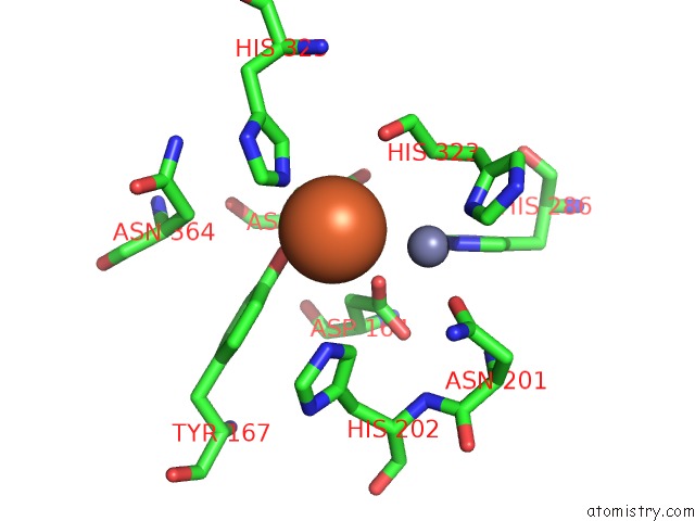
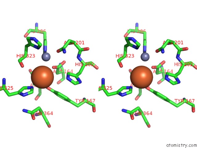
Iron binding site 1 out of 4 in 4dsy
Go back to
Iron binding site 1 out
of 4 in the Crystal Structure of Red Kidney Bean Purple Acid Phosphatase in Complex with Maybridge Fragment CC24201
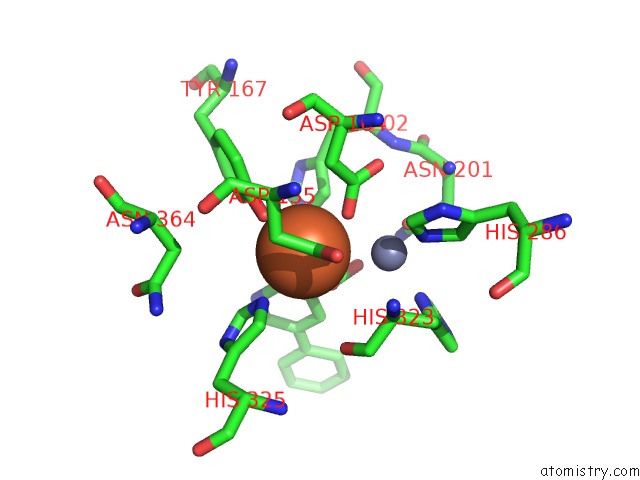
Mono view
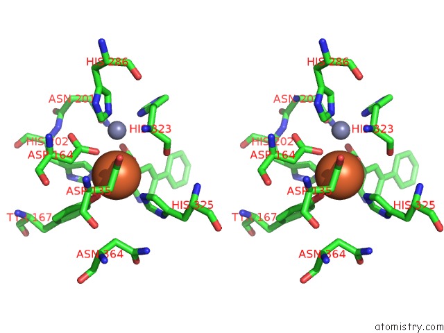
Stereo pair view
Mono view
Stereo pair view
A full contact list of Iron with other atoms in the Fe binding
site number 1 of Crystal Structure of Red Kidney Bean Purple Acid Phosphatase in Complex with Maybridge Fragment CC24201 within 5.0Å range:
|
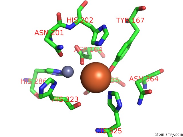
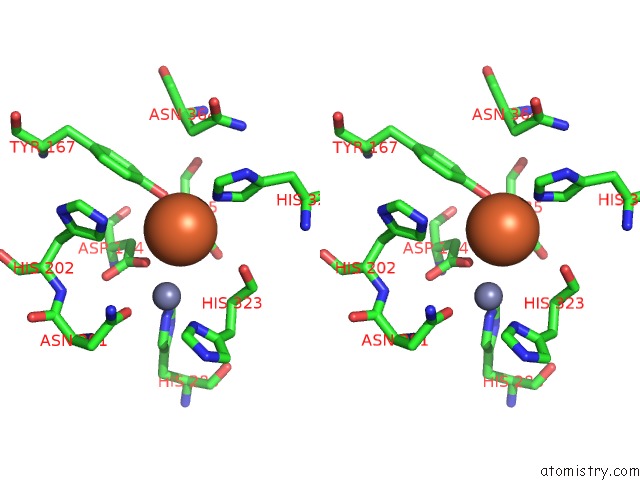
Iron binding site 2 out of 4 in 4dsy
Go back to
Iron binding site 2 out
of 4 in the Crystal Structure of Red Kidney Bean Purple Acid Phosphatase in Complex with Maybridge Fragment CC24201
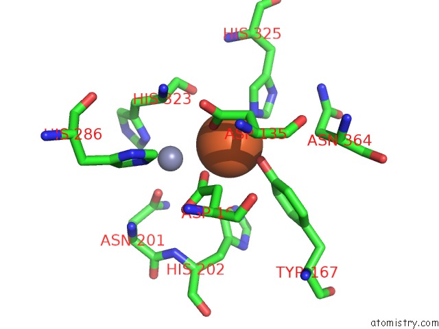
Mono view
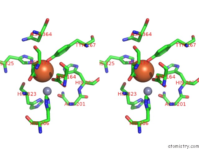
Stereo pair view
Mono view
Stereo pair view
A full contact list of Iron with other atoms in the Fe binding
site number 2 of Crystal Structure of Red Kidney Bean Purple Acid Phosphatase in Complex with Maybridge Fragment CC24201 within 5.0Å range:
|
Iron binding site 3 out of 4 in 4dsy
Go back to
Iron binding site 3 out
of 4 in the Crystal Structure of Red Kidney Bean Purple Acid Phosphatase in Complex with Maybridge Fragment CC24201
Mono view
Stereo pair view
Mono view
Stereo pair view
A full contact list of Iron with other atoms in the Fe binding
site number 3 of Crystal Structure of Red Kidney Bean Purple Acid Phosphatase in Complex with Maybridge Fragment CC24201 within 5.0Å range:
|
Iron binding site 4 out of 4 in 4dsy
Go back to
Iron binding site 4 out
of 4 in the Crystal Structure of Red Kidney Bean Purple Acid Phosphatase in Complex with Maybridge Fragment CC24201
Mono view
Stereo pair view
Mono view
Stereo pair view
A full contact list of Iron with other atoms in the Fe binding
site number 4 of Crystal Structure of Red Kidney Bean Purple Acid Phosphatase in Complex with Maybridge Fragment CC24201 within 5.0Å range:
|
Reference:
D.Feder,
W.M.Hussein,
D.J.Clayton,
M.W.Kan,
G.Schenk,
R.P.Mcgeary,
L.W.Guddat.
Identification of Purple Acid Phosphatase Inhibitors By Fragment-Based Screening: Promising New Leads For Osteoporosis Therapeutics. Chem.Biol.Drug Des. V. 80 665 2012.
ISSN: ISSN 1747-0277
PubMed: 22943065
DOI: 10.1111/CBDD.12001
Page generated: Mon Aug 5 01:20:25 2024
ISSN: ISSN 1747-0277
PubMed: 22943065
DOI: 10.1111/CBDD.12001
Last articles
Zn in 9MJ5Zn in 9HNW
Zn in 9G0L
Zn in 9FNE
Zn in 9DZN
Zn in 9E0I
Zn in 9D32
Zn in 9DAK
Zn in 8ZXC
Zn in 8ZUF