Iron »
PDB 4dig-4egm »
4dwt »
Iron in PDB 4dwt: Carbonmonoxy Dehaloperoxidase-Hemoglobin A Structure at 2.05 Angstrom Resolution
Protein crystallography data
The structure of Carbonmonoxy Dehaloperoxidase-Hemoglobin A Structure at 2.05 Angstrom Resolution, PDB code: 4dwt
was solved by
V.S.De Serrano,
J.Zhao,
S.Franzen,
with X-Ray Crystallography technique. A brief refinement statistics is given in the table below:
| Resolution Low / High (Å) | 35.00 / 2.05 |
| Space group | P 21 21 21 |
| Cell size a, b, c (Å), α, β, γ (°) | 57.910, 67.470, 68.720, 90.00, 90.00, 90.00 |
| R / Rfree (%) | 20 / 23.8 |
Iron Binding Sites:
The binding sites of Iron atom in the Carbonmonoxy Dehaloperoxidase-Hemoglobin A Structure at 2.05 Angstrom Resolution
(pdb code 4dwt). This binding sites where shown within
5.0 Angstroms radius around Iron atom.
In total 2 binding sites of Iron where determined in the Carbonmonoxy Dehaloperoxidase-Hemoglobin A Structure at 2.05 Angstrom Resolution, PDB code: 4dwt:
Jump to Iron binding site number: 1; 2;
In total 2 binding sites of Iron where determined in the Carbonmonoxy Dehaloperoxidase-Hemoglobin A Structure at 2.05 Angstrom Resolution, PDB code: 4dwt:
Jump to Iron binding site number: 1; 2;
Iron binding site 1 out of 2 in 4dwt
Go back to
Iron binding site 1 out
of 2 in the Carbonmonoxy Dehaloperoxidase-Hemoglobin A Structure at 2.05 Angstrom Resolution
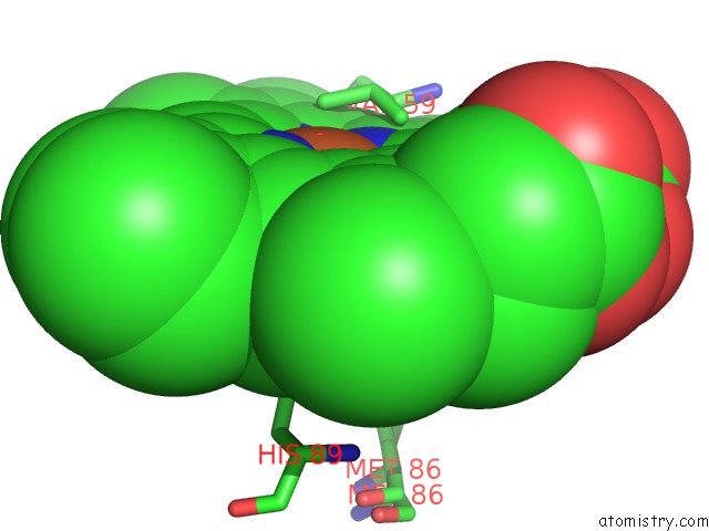
Mono view
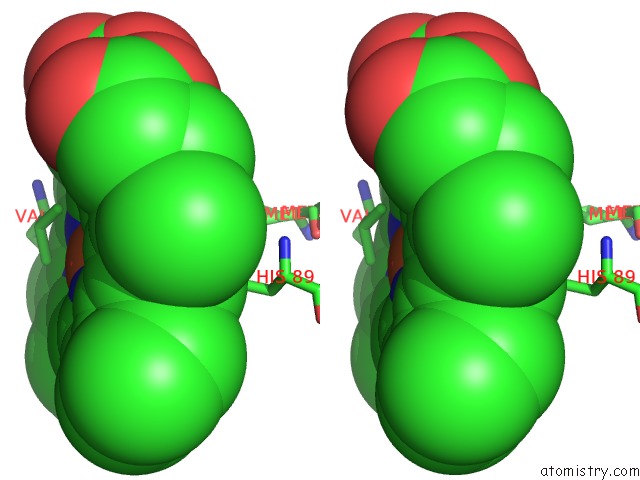
Stereo pair view
Mono view
Stereo pair view
A full contact list of Iron with other atoms in the Fe binding
site number 1 of Carbonmonoxy Dehaloperoxidase-Hemoglobin A Structure at 2.05 Angstrom Resolution within 5.0Å range:
|
Iron binding site 2 out of 2 in 4dwt
Go back to
Iron binding site 2 out
of 2 in the Carbonmonoxy Dehaloperoxidase-Hemoglobin A Structure at 2.05 Angstrom Resolution

Mono view
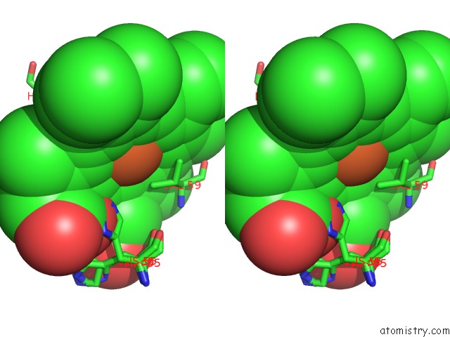
Stereo pair view
Mono view
Stereo pair view
A full contact list of Iron with other atoms in the Fe binding
site number 2 of Carbonmonoxy Dehaloperoxidase-Hemoglobin A Structure at 2.05 Angstrom Resolution within 5.0Å range:
|
Reference:
V.S.De Serrano,
J.Zhao,
S.Franzen.
A Unique Role For the Distal Histidine Observed in Carbonmonoxy Dehaloperoxidase-Hemoglobin A Structures To Be Published.
Page generated: Tue Aug 5 09:56:47 2025
Last articles
Fe in 5OBAFe in 5O31
Fe in 5OD5
Fe in 5OCF
Fe in 5OBP
Fe in 5OCB
Fe in 5OC0
Fe in 5OBI
Fe in 5OBO
Fe in 5OAY