Iron »
PDB 4egn-4f2n »
4f0l »
Iron in PDB 4f0l: Crystal Structure of Amidohydrolase From Brucella Melitensis
Protein crystallography data
The structure of Crystal Structure of Amidohydrolase From Brucella Melitensis, PDB code: 4f0l
was solved by
Seattle Structural Genomics Center For Infectious Disease (Ssgcid),
with X-Ray Crystallography technique. A brief refinement statistics is given in the table below:
| Resolution Low / High (Å) | 50.00 / 2.05 |
| Space group | P 1 |
| Cell size a, b, c (Å), α, β, γ (°) | 53.592, 62.192, 68.926, 92.75, 95.49, 112.32 |
| R / Rfree (%) | 14.8 / 18.4 |
Iron Binding Sites:
The binding sites of Iron atom in the Crystal Structure of Amidohydrolase From Brucella Melitensis
(pdb code 4f0l). This binding sites where shown within
5.0 Angstroms radius around Iron atom.
In total 2 binding sites of Iron where determined in the Crystal Structure of Amidohydrolase From Brucella Melitensis, PDB code: 4f0l:
Jump to Iron binding site number: 1; 2;
In total 2 binding sites of Iron where determined in the Crystal Structure of Amidohydrolase From Brucella Melitensis, PDB code: 4f0l:
Jump to Iron binding site number: 1; 2;
Iron binding site 1 out of 2 in 4f0l
Go back to
Iron binding site 1 out
of 2 in the Crystal Structure of Amidohydrolase From Brucella Melitensis
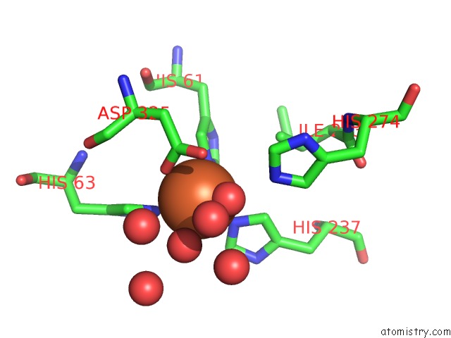
Mono view
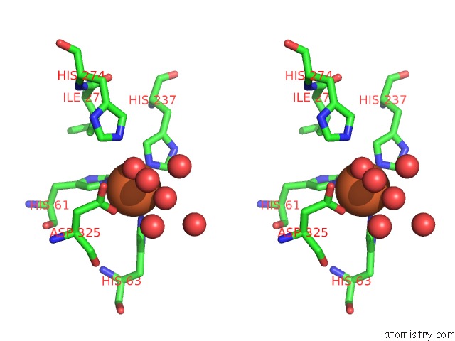
Stereo pair view
Mono view
Stereo pair view
A full contact list of Iron with other atoms in the Fe binding
site number 1 of Crystal Structure of Amidohydrolase From Brucella Melitensis within 5.0Å range:
|
Iron binding site 2 out of 2 in 4f0l
Go back to
Iron binding site 2 out
of 2 in the Crystal Structure of Amidohydrolase From Brucella Melitensis
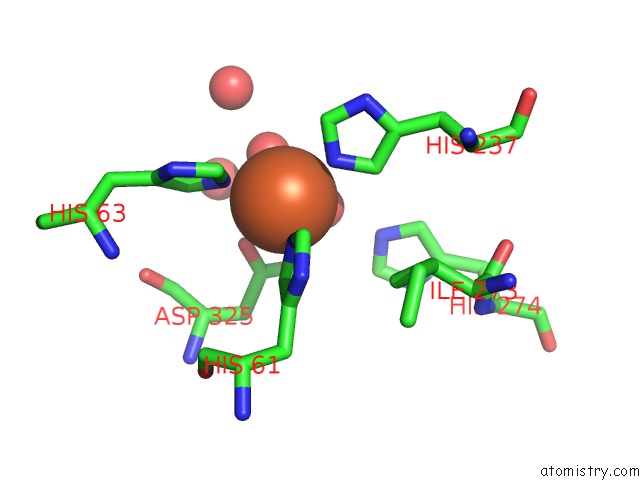
Mono view
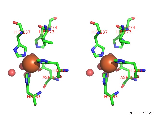
Stereo pair view
Mono view
Stereo pair view
A full contact list of Iron with other atoms in the Fe binding
site number 2 of Crystal Structure of Amidohydrolase From Brucella Melitensis within 5.0Å range:
|
Reference:
A.S.Gardberg,
J.Abendroth,
B.Staker,
L.Stewart,
Seattle Structural Genomics Center For Infectious Disease(Ssgcid).
Crystal Structure of Amidohydrolase From Brucella Melitensis To Be Published.
Page generated: Tue Aug 5 10:11:38 2025
Last articles
Fe in 4O2GFe in 4O1Q
Fe in 4NZI
Fe in 4O1T
Fe in 4NZ2
Fe in 4NY4
Fe in 4NXC
Fe in 4NXA
Fe in 4NVO
Fe in 4NVN