Iron »
PDB 4f2z-4fh6 »
4f2z »
Iron in PDB 4f2z: Crystal Structure of RPE65 in A Lipid Environment
Enzymatic activity of Crystal Structure of RPE65 in A Lipid Environment
All present enzymatic activity of Crystal Structure of RPE65 in A Lipid Environment:
3.1.1.64;
3.1.1.64;
Protein crystallography data
The structure of Crystal Structure of RPE65 in A Lipid Environment, PDB code: 4f2z
was solved by
P.D.Kiser,
W.Shi,
K.Palczewski,
with X-Ray Crystallography technique. A brief refinement statistics is given in the table below:
| Resolution Low / High (Å) | 69.40 / 3.00 |
| Space group | P 21 21 21 |
| Cell size a, b, c (Å), α, β, γ (°) | 55.799, 100.234, 277.588, 90.00, 90.00, 90.00 |
| R / Rfree (%) | 22.4 / 26.1 |
Iron Binding Sites:
The binding sites of Iron atom in the Crystal Structure of RPE65 in A Lipid Environment
(pdb code 4f2z). This binding sites where shown within
5.0 Angstroms radius around Iron atom.
In total 2 binding sites of Iron where determined in the Crystal Structure of RPE65 in A Lipid Environment, PDB code: 4f2z:
Jump to Iron binding site number: 1; 2;
In total 2 binding sites of Iron where determined in the Crystal Structure of RPE65 in A Lipid Environment, PDB code: 4f2z:
Jump to Iron binding site number: 1; 2;
Iron binding site 1 out of 2 in 4f2z
Go back to
Iron binding site 1 out
of 2 in the Crystal Structure of RPE65 in A Lipid Environment
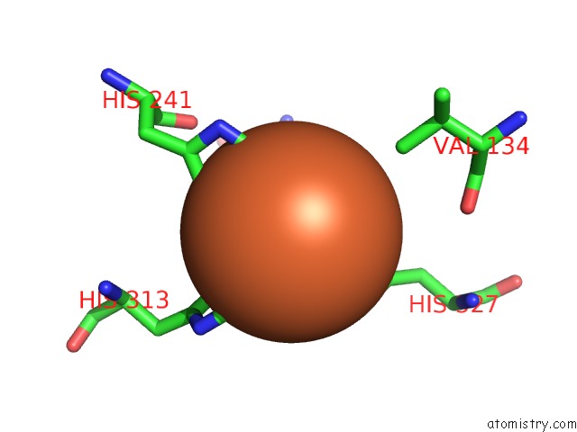
Mono view
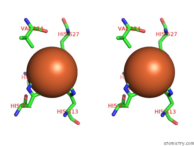
Stereo pair view
Mono view
Stereo pair view
A full contact list of Iron with other atoms in the Fe binding
site number 1 of Crystal Structure of RPE65 in A Lipid Environment within 5.0Å range:
|
Iron binding site 2 out of 2 in 4f2z
Go back to
Iron binding site 2 out
of 2 in the Crystal Structure of RPE65 in A Lipid Environment
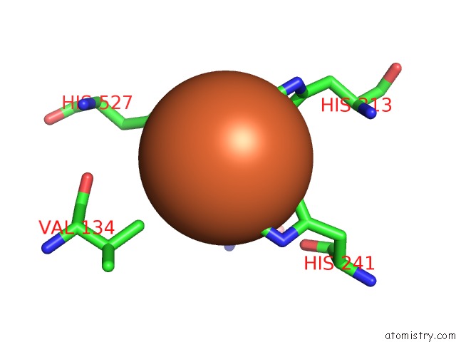
Mono view
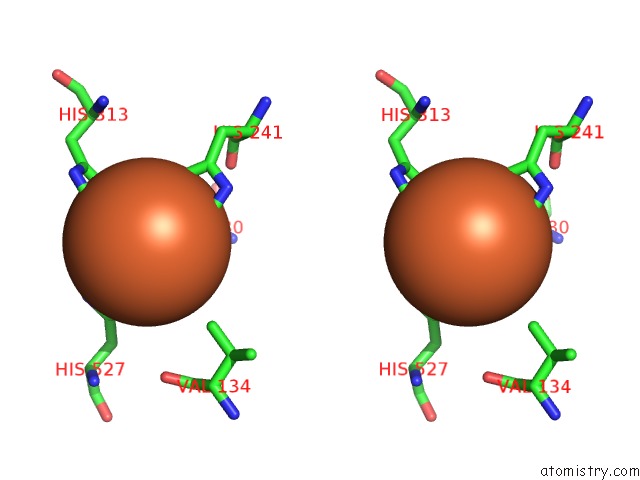
Stereo pair view
Mono view
Stereo pair view
A full contact list of Iron with other atoms in the Fe binding
site number 2 of Crystal Structure of RPE65 in A Lipid Environment within 5.0Å range:
|
Reference:
P.D.Kiser,
E.R.Farquhar,
W.Shi,
X.Sui,
M.R.Chance,
K.Palczewski.
Structure of RPE65 Isomerase in A Lipidic Matrix Reveals Roles For Phospholipids and Iron in Catalysis. Proc.Natl.Acad.Sci.Usa V. 109 E2747 2012.
ISSN: ISSN 0027-8424
PubMed: 23012475
DOI: 10.1073/PNAS.1212025109
Page generated: Mon Aug 5 02:05:31 2024
ISSN: ISSN 0027-8424
PubMed: 23012475
DOI: 10.1073/PNAS.1212025109
Last articles
Zn in 9MJ5Zn in 9HNW
Zn in 9G0L
Zn in 9FNE
Zn in 9DZN
Zn in 9E0I
Zn in 9D32
Zn in 9DAK
Zn in 8ZXC
Zn in 8ZUF