Iron »
PDB 4f2z-4fh6 »
4fcs »
Iron in PDB 4fcs: The Crystal Structures of Several Mutants of Pleurotus Eryngii Versatile Peroxidase
Enzymatic activity of The Crystal Structures of Several Mutants of Pleurotus Eryngii Versatile Peroxidase
All present enzymatic activity of The Crystal Structures of Several Mutants of Pleurotus Eryngii Versatile Peroxidase:
1.11.1.16;
1.11.1.16;
Protein crystallography data
The structure of The Crystal Structures of Several Mutants of Pleurotus Eryngii Versatile Peroxidase, PDB code: 4fcs
was solved by
M.J.Mate,
A.Romero,
F.J.Ruiz-Duenas,
A.T.Martinez,
with X-Ray Crystallography technique. A brief refinement statistics is given in the table below:
| Resolution Low / High (Å) | 48.14 / 1.50 |
| Space group | I 41 |
| Cell size a, b, c (Å), α, β, γ (°) | 96.271, 96.271, 98.907, 90.00, 90.00, 90.00 |
| R / Rfree (%) | 15 / 17.6 |
Other elements in 4fcs:
The structure of The Crystal Structures of Several Mutants of Pleurotus Eryngii Versatile Peroxidase also contains other interesting chemical elements:
| Calcium | (Ca) | 2 atoms |
Iron Binding Sites:
The binding sites of Iron atom in the The Crystal Structures of Several Mutants of Pleurotus Eryngii Versatile Peroxidase
(pdb code 4fcs). This binding sites where shown within
5.0 Angstroms radius around Iron atom.
In total only one binding site of Iron was determined in the The Crystal Structures of Several Mutants of Pleurotus Eryngii Versatile Peroxidase, PDB code: 4fcs:
In total only one binding site of Iron was determined in the The Crystal Structures of Several Mutants of Pleurotus Eryngii Versatile Peroxidase, PDB code: 4fcs:
Iron binding site 1 out of 1 in 4fcs
Go back to
Iron binding site 1 out
of 1 in the The Crystal Structures of Several Mutants of Pleurotus Eryngii Versatile Peroxidase
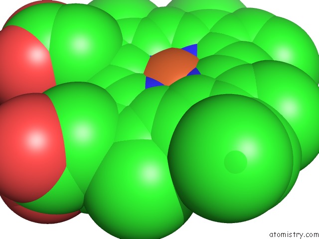
Mono view
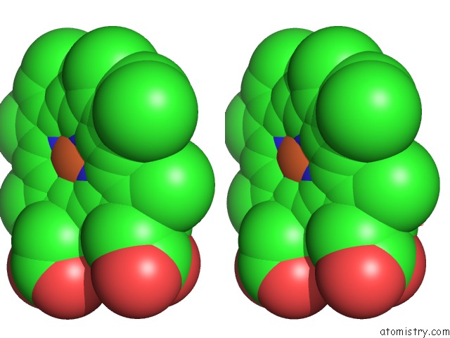
Stereo pair view
Mono view
Stereo pair view
A full contact list of Iron with other atoms in the Fe binding
site number 1 of The Crystal Structures of Several Mutants of Pleurotus Eryngii Versatile Peroxidase within 5.0Å range:
|
Reference:
M.Morales,
M.J.Mate,
A.Romero,
M.J.Martinez,
A.T.Martinez,
F.J.Ruiz-Duenas.
Two Oxidation Sites For Low Redox Potential Substrates: A Directed Mutagenesis, Kinetic, and Crystallographic Study on Pleurotus Eryngii Versatile Peroxidase. J.Biol.Chem. V. 287 41053 2012.
ISSN: ISSN 0021-9258
PubMed: 23071108
DOI: 10.1074/JBC.M112.405548
Page generated: Mon Aug 5 02:12:20 2024
ISSN: ISSN 0021-9258
PubMed: 23071108
DOI: 10.1074/JBC.M112.405548
Last articles
Zn in 9JYWZn in 9IR4
Zn in 9IR3
Zn in 9GMX
Zn in 9GMW
Zn in 9JEJ
Zn in 9ERF
Zn in 9ERE
Zn in 9EGV
Zn in 9EGW