Iron »
PDB 4g39-4gl5 »
4g7s »
Iron in PDB 4g7s: Structure of Recombinant Cytochrome BA3 Oxidase Mutant V236I From Thermus Thermophilus
Enzymatic activity of Structure of Recombinant Cytochrome BA3 Oxidase Mutant V236I From Thermus Thermophilus
All present enzymatic activity of Structure of Recombinant Cytochrome BA3 Oxidase Mutant V236I From Thermus Thermophilus:
1.9.3.1;
1.9.3.1;
Protein crystallography data
The structure of Structure of Recombinant Cytochrome BA3 Oxidase Mutant V236I From Thermus Thermophilus, PDB code: 4g7s
was solved by
Y.Li,
Y.Chen,
C.D.Stout,
with X-Ray Crystallography technique. A brief refinement statistics is given in the table below:
| Resolution Low / High (Å) | 74.89 / 2.00 |
| Space group | C 1 2 1 |
| Cell size a, b, c (Å), α, β, γ (°) | 143.570, 98.390, 94.580, 90.00, 127.64, 90.00 |
| R / Rfree (%) | 18.2 / 20.7 |
Other elements in 4g7s:
The structure of Structure of Recombinant Cytochrome BA3 Oxidase Mutant V236I From Thermus Thermophilus also contains other interesting chemical elements:
| Copper | (Cu) | 3 atoms |
Iron Binding Sites:
The binding sites of Iron atom in the Structure of Recombinant Cytochrome BA3 Oxidase Mutant V236I From Thermus Thermophilus
(pdb code 4g7s). This binding sites where shown within
5.0 Angstroms radius around Iron atom.
In total 2 binding sites of Iron where determined in the Structure of Recombinant Cytochrome BA3 Oxidase Mutant V236I From Thermus Thermophilus, PDB code: 4g7s:
Jump to Iron binding site number: 1; 2;
In total 2 binding sites of Iron where determined in the Structure of Recombinant Cytochrome BA3 Oxidase Mutant V236I From Thermus Thermophilus, PDB code: 4g7s:
Jump to Iron binding site number: 1; 2;
Iron binding site 1 out of 2 in 4g7s
Go back to
Iron binding site 1 out
of 2 in the Structure of Recombinant Cytochrome BA3 Oxidase Mutant V236I From Thermus Thermophilus
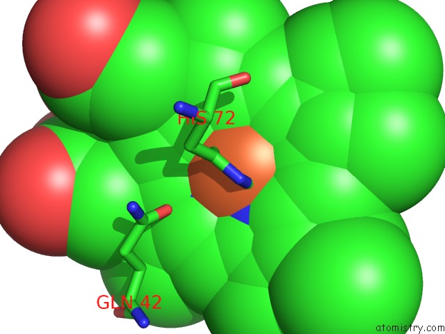
Mono view
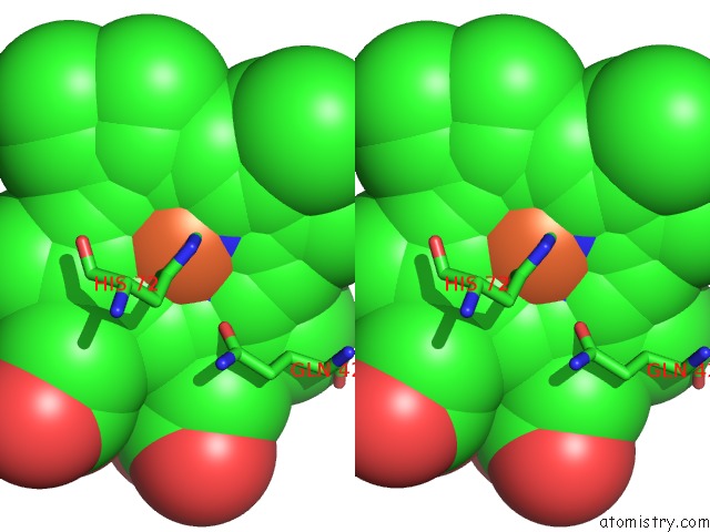
Stereo pair view
Mono view
Stereo pair view
A full contact list of Iron with other atoms in the Fe binding
site number 1 of Structure of Recombinant Cytochrome BA3 Oxidase Mutant V236I From Thermus Thermophilus within 5.0Å range:
|
Iron binding site 2 out of 2 in 4g7s
Go back to
Iron binding site 2 out
of 2 in the Structure of Recombinant Cytochrome BA3 Oxidase Mutant V236I From Thermus Thermophilus
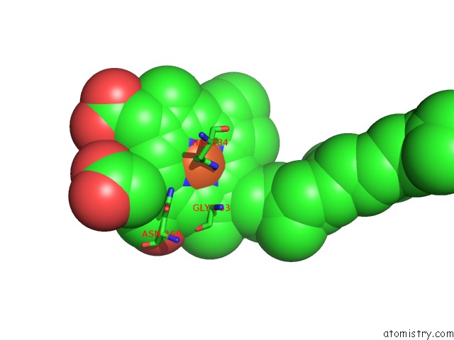
Mono view
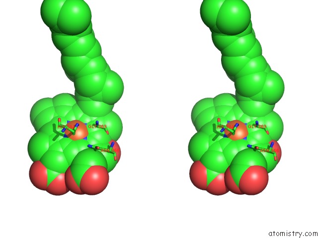
Stereo pair view
Mono view
Stereo pair view
A full contact list of Iron with other atoms in the Fe binding
site number 2 of Structure of Recombinant Cytochrome BA3 Oxidase Mutant V236I From Thermus Thermophilus within 5.0Å range:
|
Reference:
Y.Li,
Y.Chen,
C.D.Stout.
Structure of Recombinant Cytochrome BA3 Oxidase Mutant V236I From Thermus Thermophilus To Be Published.
Page generated: Mon Aug 5 02:39:16 2024
Last articles
Fe in 2YXOFe in 2YRS
Fe in 2YXC
Fe in 2YNM
Fe in 2YVJ
Fe in 2YP1
Fe in 2YU2
Fe in 2YU1
Fe in 2YQB
Fe in 2YOO