Iron »
PDB 4g39-4gl5 »
4gam »
Iron in PDB 4gam: Complex Structure of Methane Monooxygenase Hydroxylase and Regulatory Subunit
Enzymatic activity of Complex Structure of Methane Monooxygenase Hydroxylase and Regulatory Subunit
All present enzymatic activity of Complex Structure of Methane Monooxygenase Hydroxylase and Regulatory Subunit:
1.14.13.25;
1.14.13.25;
Protein crystallography data
The structure of Complex Structure of Methane Monooxygenase Hydroxylase and Regulatory Subunit, PDB code: 4gam
was solved by
S.J.Lee,
S.J.Lippard,
U.-S.Cho,
with X-Ray Crystallography technique. A brief refinement statistics is given in the table below:
| Resolution Low / High (Å) | 50.00 / 2.90 |
| Space group | P 21 21 21 |
| Cell size a, b, c (Å), α, β, γ (°) | 183.590, 248.970, 122.290, 90.00, 90.00, 90.00 |
| R / Rfree (%) | 20.5 / 25.8 |
Iron Binding Sites:
The binding sites of Iron atom in the Complex Structure of Methane Monooxygenase Hydroxylase and Regulatory Subunit
(pdb code 4gam). This binding sites where shown within
5.0 Angstroms radius around Iron atom.
In total 8 binding sites of Iron where determined in the Complex Structure of Methane Monooxygenase Hydroxylase and Regulatory Subunit, PDB code: 4gam:
Jump to Iron binding site number: 1; 2; 3; 4; 5; 6; 7; 8;
In total 8 binding sites of Iron where determined in the Complex Structure of Methane Monooxygenase Hydroxylase and Regulatory Subunit, PDB code: 4gam:
Jump to Iron binding site number: 1; 2; 3; 4; 5; 6; 7; 8;
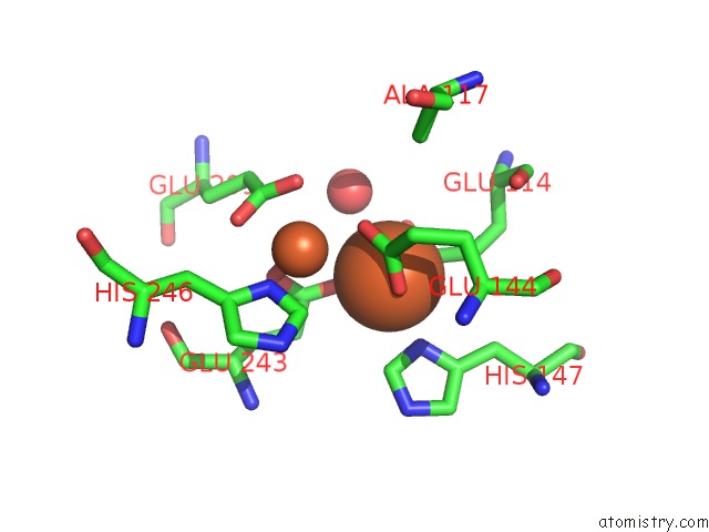
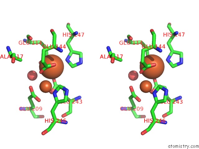
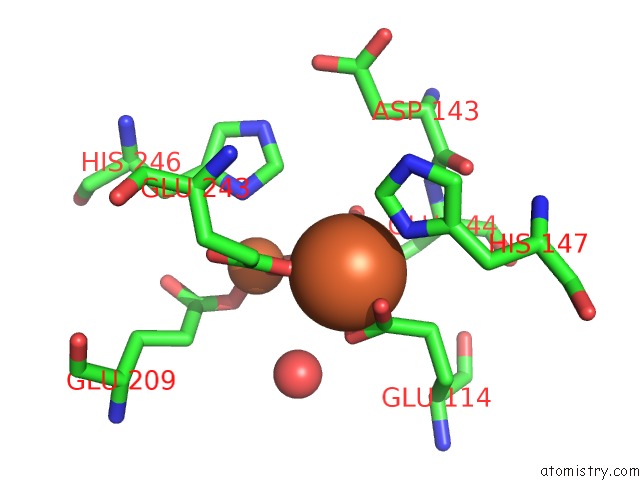
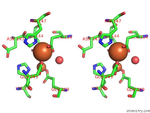
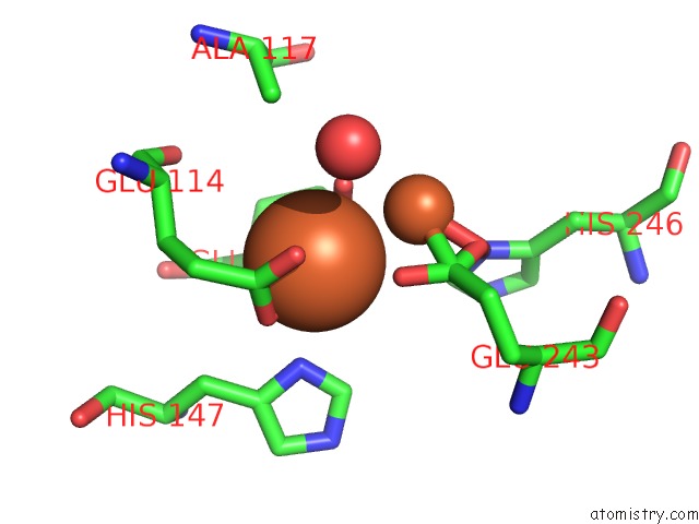
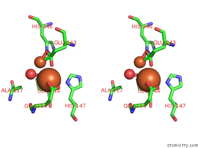
Iron binding site 1 out of 8 in 4gam
Go back to
Iron binding site 1 out
of 8 in the Complex Structure of Methane Monooxygenase Hydroxylase and Regulatory Subunit
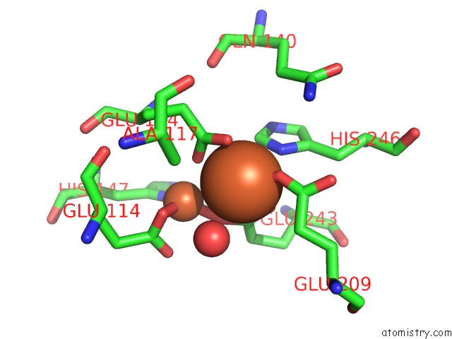
Mono view
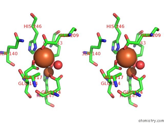
Stereo pair view
Mono view
Stereo pair view
A full contact list of Iron with other atoms in the Fe binding
site number 1 of Complex Structure of Methane Monooxygenase Hydroxylase and Regulatory Subunit within 5.0Å range:
|
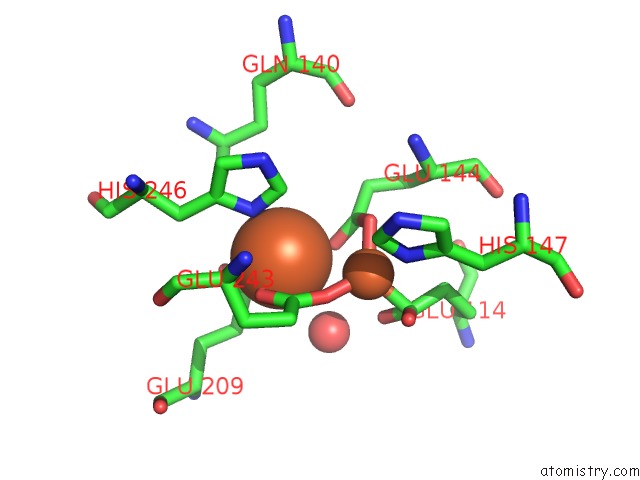
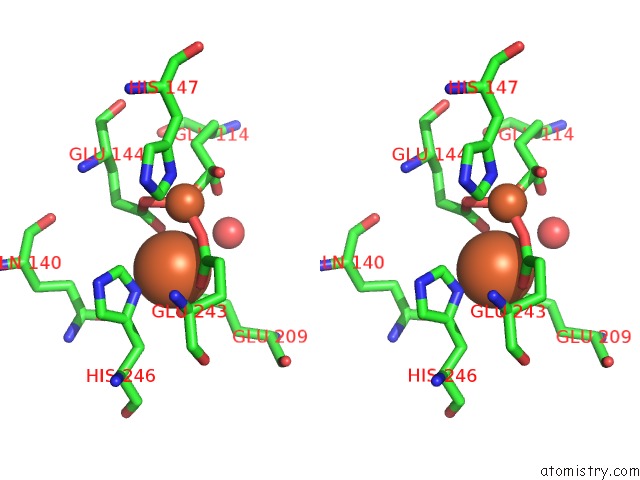
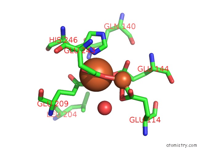
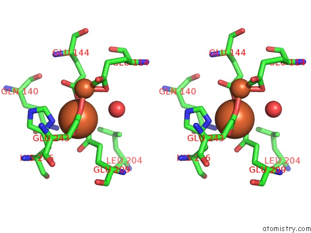
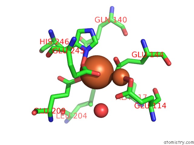
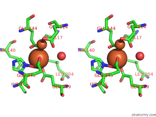
Iron binding site 2 out of 8 in 4gam
Go back to
Iron binding site 2 out
of 8 in the Complex Structure of Methane Monooxygenase Hydroxylase and Regulatory Subunit
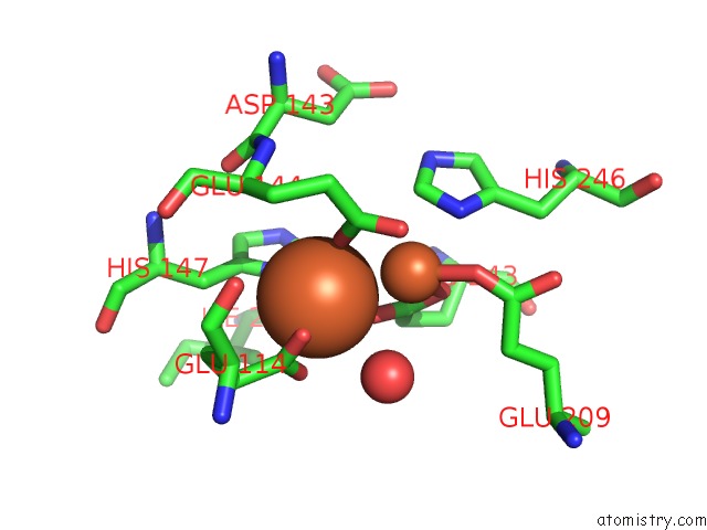
Mono view
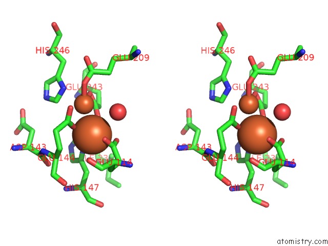
Stereo pair view
Mono view
Stereo pair view
A full contact list of Iron with other atoms in the Fe binding
site number 2 of Complex Structure of Methane Monooxygenase Hydroxylase and Regulatory Subunit within 5.0Å range:
|
Iron binding site 3 out of 8 in 4gam
Go back to
Iron binding site 3 out
of 8 in the Complex Structure of Methane Monooxygenase Hydroxylase and Regulatory Subunit
Mono view
Stereo pair view
Mono view
Stereo pair view
A full contact list of Iron with other atoms in the Fe binding
site number 3 of Complex Structure of Methane Monooxygenase Hydroxylase and Regulatory Subunit within 5.0Å range:
|
Iron binding site 4 out of 8 in 4gam
Go back to
Iron binding site 4 out
of 8 in the Complex Structure of Methane Monooxygenase Hydroxylase and Regulatory Subunit
Mono view
Stereo pair view
Mono view
Stereo pair view
A full contact list of Iron with other atoms in the Fe binding
site number 4 of Complex Structure of Methane Monooxygenase Hydroxylase and Regulatory Subunit within 5.0Å range:
|
Iron binding site 5 out of 8 in 4gam
Go back to
Iron binding site 5 out
of 8 in the Complex Structure of Methane Monooxygenase Hydroxylase and Regulatory Subunit
Mono view
Stereo pair view
Mono view
Stereo pair view
A full contact list of Iron with other atoms in the Fe binding
site number 5 of Complex Structure of Methane Monooxygenase Hydroxylase and Regulatory Subunit within 5.0Å range:
|
Iron binding site 6 out of 8 in 4gam
Go back to
Iron binding site 6 out
of 8 in the Complex Structure of Methane Monooxygenase Hydroxylase and Regulatory Subunit
Mono view
Stereo pair view
Mono view
Stereo pair view
A full contact list of Iron with other atoms in the Fe binding
site number 6 of Complex Structure of Methane Monooxygenase Hydroxylase and Regulatory Subunit within 5.0Å range:
|
Iron binding site 7 out of 8 in 4gam
Go back to
Iron binding site 7 out
of 8 in the Complex Structure of Methane Monooxygenase Hydroxylase and Regulatory Subunit
Mono view
Stereo pair view
Mono view
Stereo pair view
A full contact list of Iron with other atoms in the Fe binding
site number 7 of Complex Structure of Methane Monooxygenase Hydroxylase and Regulatory Subunit within 5.0Å range:
|
Iron binding site 8 out of 8 in 4gam
Go back to
Iron binding site 8 out
of 8 in the Complex Structure of Methane Monooxygenase Hydroxylase and Regulatory Subunit
Mono view
Stereo pair view
Mono view
Stereo pair view
A full contact list of Iron with other atoms in the Fe binding
site number 8 of Complex Structure of Methane Monooxygenase Hydroxylase and Regulatory Subunit within 5.0Å range:
|
Reference:
S.J.Lee,
M.S.Mccormick,
S.J.Lippard,
U.S.Cho.
Control of Substrate Access to the Active Site in Methane Monooxygenase. Nature V. 494 380 2013.
ISSN: ISSN 0028-0836
PubMed: 23395959
DOI: 10.1038/NATURE11880
Page generated: Tue Aug 5 10:36:40 2025
ISSN: ISSN 0028-0836
PubMed: 23395959
DOI: 10.1038/NATURE11880
Last articles
Fe in 5TH5Fe in 5T5M
Fe in 5TIA
Fe in 5TI9
Fe in 5TGS
Fe in 5TFU
Fe in 5TG0
Fe in 5TFT
Fe in 5TE8
Fe in 5TDV