Iron »
PDB 4g39-4gl5 »
4ghd »
Iron in PDB 4ghd: Structure of Y257F Variant of Homoprotocatechuate 2,3-Dioxygenase From B.Fuscum in Complex with Hpca at 1.85 Ang Resolution
Enzymatic activity of Structure of Y257F Variant of Homoprotocatechuate 2,3-Dioxygenase From B.Fuscum in Complex with Hpca at 1.85 Ang Resolution
All present enzymatic activity of Structure of Y257F Variant of Homoprotocatechuate 2,3-Dioxygenase From B.Fuscum in Complex with Hpca at 1.85 Ang Resolution:
1.13.11.15;
1.13.11.15;
Protein crystallography data
The structure of Structure of Y257F Variant of Homoprotocatechuate 2,3-Dioxygenase From B.Fuscum in Complex with Hpca at 1.85 Ang Resolution, PDB code: 4ghd
was solved by
E.G.Kovaleva,
J.D.Lipscomb,
with X-Ray Crystallography technique. A brief refinement statistics is given in the table below:
| Resolution Low / High (Å) | 48.05 / 1.85 |
| Space group | P 21 21 2 |
| Cell size a, b, c (Å), α, β, γ (°) | 110.163, 150.701, 96.107, 90.00, 90.00, 90.00 |
| R / Rfree (%) | 15.2 / 18.9 |
Other elements in 4ghd:
The structure of Structure of Y257F Variant of Homoprotocatechuate 2,3-Dioxygenase From B.Fuscum in Complex with Hpca at 1.85 Ang Resolution also contains other interesting chemical elements:
| Chlorine | (Cl) | 3 atoms |
| Calcium | (Ca) | 1 atom |
Iron Binding Sites:
The binding sites of Iron atom in the Structure of Y257F Variant of Homoprotocatechuate 2,3-Dioxygenase From B.Fuscum in Complex with Hpca at 1.85 Ang Resolution
(pdb code 4ghd). This binding sites where shown within
5.0 Angstroms radius around Iron atom.
In total 4 binding sites of Iron where determined in the Structure of Y257F Variant of Homoprotocatechuate 2,3-Dioxygenase From B.Fuscum in Complex with Hpca at 1.85 Ang Resolution, PDB code: 4ghd:
Jump to Iron binding site number: 1; 2; 3; 4;
In total 4 binding sites of Iron where determined in the Structure of Y257F Variant of Homoprotocatechuate 2,3-Dioxygenase From B.Fuscum in Complex with Hpca at 1.85 Ang Resolution, PDB code: 4ghd:
Jump to Iron binding site number: 1; 2; 3; 4;
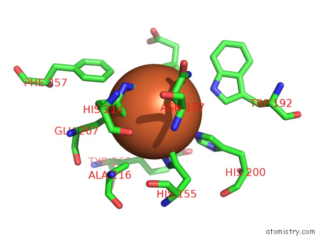
Iron binding site 1 out of 4 in 4ghd
Go back to
Iron binding site 1 out
of 4 in the Structure of Y257F Variant of Homoprotocatechuate 2,3-Dioxygenase From B.Fuscum in Complex with Hpca at 1.85 Ang Resolution
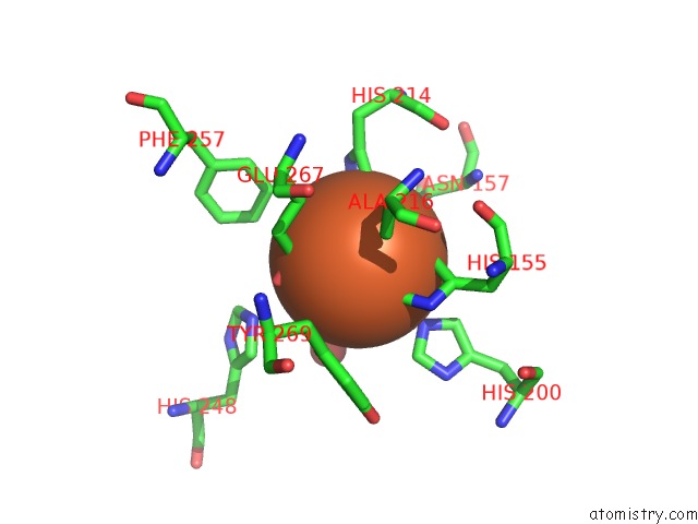
Mono view
Stereo pair view
Mono view
Stereo pair view
A full contact list of Iron with other atoms in the Fe binding
site number 1 of Structure of Y257F Variant of Homoprotocatechuate 2,3-Dioxygenase From B.Fuscum in Complex with Hpca at 1.85 Ang Resolution within 5.0Å range:
|
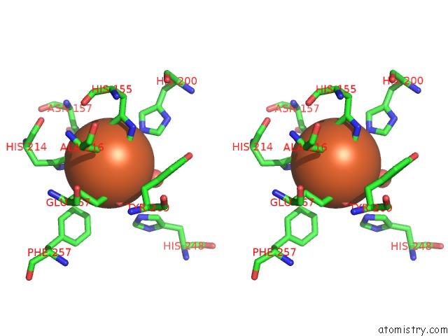
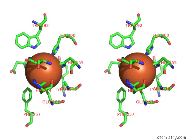
Iron binding site 2 out of 4 in 4ghd
Go back to
Iron binding site 2 out
of 4 in the Structure of Y257F Variant of Homoprotocatechuate 2,3-Dioxygenase From B.Fuscum in Complex with Hpca at 1.85 Ang Resolution
Mono view
Stereo pair view
Mono view
Stereo pair view
A full contact list of Iron with other atoms in the Fe binding
site number 2 of Structure of Y257F Variant of Homoprotocatechuate 2,3-Dioxygenase From B.Fuscum in Complex with Hpca at 1.85 Ang Resolution within 5.0Å range:
|
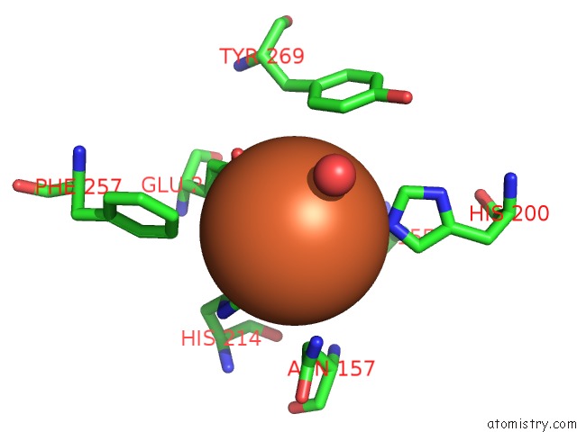
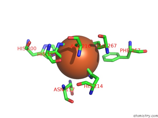
Iron binding site 3 out of 4 in 4ghd
Go back to
Iron binding site 3 out
of 4 in the Structure of Y257F Variant of Homoprotocatechuate 2,3-Dioxygenase From B.Fuscum in Complex with Hpca at 1.85 Ang Resolution
Mono view
Stereo pair view
Mono view
Stereo pair view
A full contact list of Iron with other atoms in the Fe binding
site number 3 of Structure of Y257F Variant of Homoprotocatechuate 2,3-Dioxygenase From B.Fuscum in Complex with Hpca at 1.85 Ang Resolution within 5.0Å range:
|
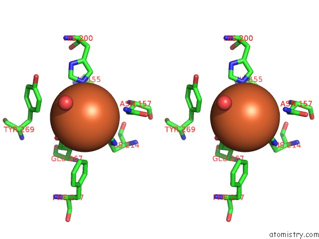
Iron binding site 4 out of 4 in 4ghd
Go back to
Iron binding site 4 out
of 4 in the Structure of Y257F Variant of Homoprotocatechuate 2,3-Dioxygenase From B.Fuscum in Complex with Hpca at 1.85 Ang Resolution
Mono view
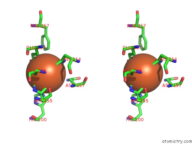
Stereo pair view
Mono view
Stereo pair view
A full contact list of Iron with other atoms in the Fe binding
site number 4 of Structure of Y257F Variant of Homoprotocatechuate 2,3-Dioxygenase From B.Fuscum in Complex with Hpca at 1.85 Ang Resolution within 5.0Å range:
|
Reference:
E.G.Kovaleva,
J.D.Lipscomb.
Structural Basis For the Role of Tyrosine 257 of Homoprotocatechuate 2,3-Dioxygenase in Substrate and Oxygen Activation. Biochemistry V. 51 8755 2012.
ISSN: ISSN 0006-2960
PubMed: 23066739
DOI: 10.1021/BI301115C
Page generated: Mon Aug 5 02:42:24 2024
ISSN: ISSN 0006-2960
PubMed: 23066739
DOI: 10.1021/BI301115C
Last articles
Cl in 5UN3Cl in 5UOA
Cl in 5UNN
Cl in 5UMH
Cl in 5UMM
Cl in 5UM6
Cl in 5UM1
Cl in 5ULX
Cl in 5ULT
Cl in 5UKA