Iron »
PDB 4gl7-4h9l »
4gqs »
Iron in PDB 4gqs: Structure of Human Microsomal Cytochrome P450 (Cyp) 2C19
Enzymatic activity of Structure of Human Microsomal Cytochrome P450 (Cyp) 2C19
All present enzymatic activity of Structure of Human Microsomal Cytochrome P450 (Cyp) 2C19:
1.14.13.48; 1.14.13.49; 1.14.13.80;
1.14.13.48; 1.14.13.49; 1.14.13.80;
Protein crystallography data
The structure of Structure of Human Microsomal Cytochrome P450 (Cyp) 2C19, PDB code: 4gqs
was solved by
R.L.Reynald,
S.Sansen,
C.D.Stout,
E.F.Johnson,
with X-Ray Crystallography technique. A brief refinement statistics is given in the table below:
| Resolution Low / High (Å) | 79.60 / 2.87 |
| Space group | H 3 2 |
| Cell size a, b, c (Å), α, β, γ (°) | 159.189, 159.189, 450.027, 90.00, 90.00, 120.00 |
| R / Rfree (%) | 25 / 29.6 |
Iron Binding Sites:
The binding sites of Iron atom in the Structure of Human Microsomal Cytochrome P450 (Cyp) 2C19
(pdb code 4gqs). This binding sites where shown within
5.0 Angstroms radius around Iron atom.
In total 4 binding sites of Iron where determined in the Structure of Human Microsomal Cytochrome P450 (Cyp) 2C19, PDB code: 4gqs:
Jump to Iron binding site number: 1; 2; 3; 4;
In total 4 binding sites of Iron where determined in the Structure of Human Microsomal Cytochrome P450 (Cyp) 2C19, PDB code: 4gqs:
Jump to Iron binding site number: 1; 2; 3; 4;
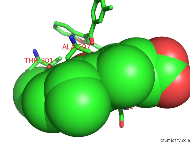
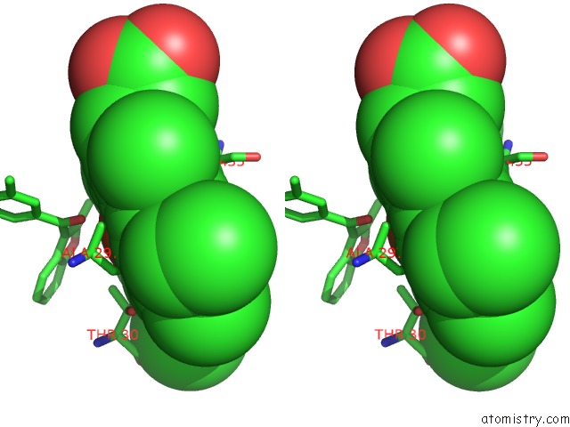
Iron binding site 1 out of 4 in 4gqs
Go back to
Iron binding site 1 out
of 4 in the Structure of Human Microsomal Cytochrome P450 (Cyp) 2C19
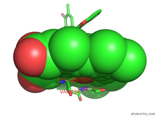
Mono view
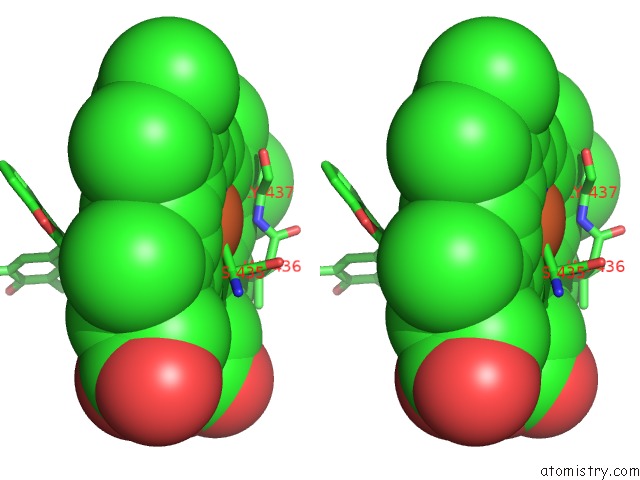
Stereo pair view
Mono view
Stereo pair view
A full contact list of Iron with other atoms in the Fe binding
site number 1 of Structure of Human Microsomal Cytochrome P450 (Cyp) 2C19 within 5.0Å range:
|
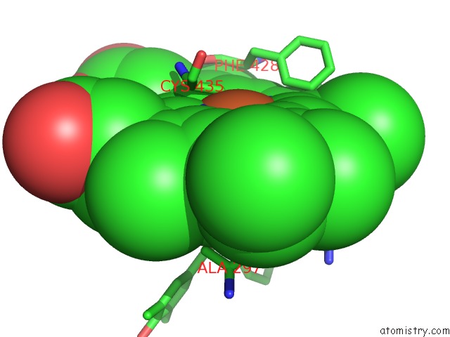
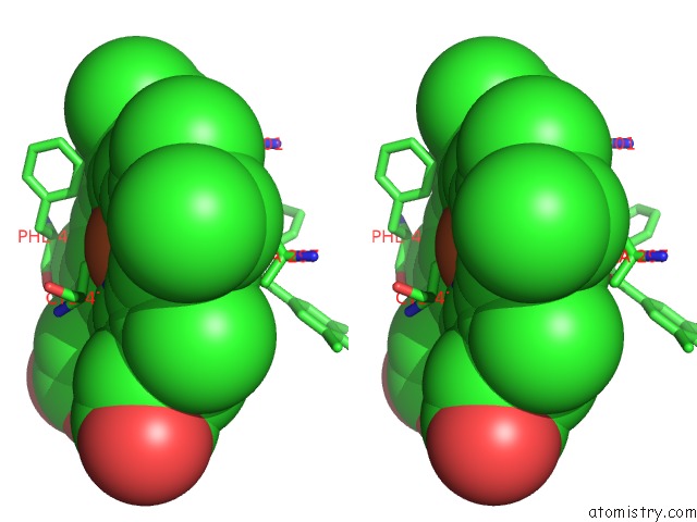
Iron binding site 2 out of 4 in 4gqs
Go back to
Iron binding site 2 out
of 4 in the Structure of Human Microsomal Cytochrome P450 (Cyp) 2C19
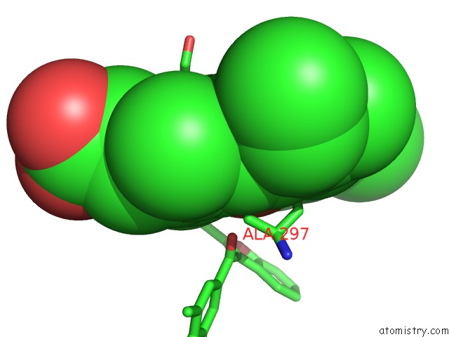
Mono view
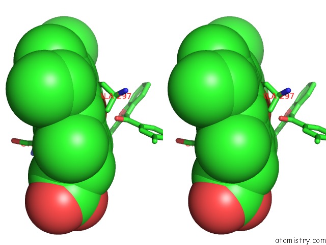
Stereo pair view
Mono view
Stereo pair view
A full contact list of Iron with other atoms in the Fe binding
site number 2 of Structure of Human Microsomal Cytochrome P450 (Cyp) 2C19 within 5.0Å range:
|
Iron binding site 3 out of 4 in 4gqs
Go back to
Iron binding site 3 out
of 4 in the Structure of Human Microsomal Cytochrome P450 (Cyp) 2C19
Mono view
Stereo pair view
Mono view
Stereo pair view
A full contact list of Iron with other atoms in the Fe binding
site number 3 of Structure of Human Microsomal Cytochrome P450 (Cyp) 2C19 within 5.0Å range:
|
Iron binding site 4 out of 4 in 4gqs
Go back to
Iron binding site 4 out
of 4 in the Structure of Human Microsomal Cytochrome P450 (Cyp) 2C19
Mono view
Stereo pair view
Mono view
Stereo pair view
A full contact list of Iron with other atoms in the Fe binding
site number 4 of Structure of Human Microsomal Cytochrome P450 (Cyp) 2C19 within 5.0Å range:
|
Reference:
R.L.Reynald,
S.Sansen,
C.D.Stout,
E.F.Johnson.
Structural Characterization of Human Cytochrome P450 2C19: Active Site Differences Between P450S 2C8, 2C9, and 2C19. J.Biol.Chem. V. 287 44581 2012.
ISSN: ISSN 0021-9258
PubMed: 23118231
DOI: 10.1074/JBC.M112.424895
Page generated: Mon Aug 5 03:02:27 2024
ISSN: ISSN 0021-9258
PubMed: 23118231
DOI: 10.1074/JBC.M112.424895
Last articles
Fe in 2YXOFe in 2YRS
Fe in 2YXC
Fe in 2YNM
Fe in 2YVJ
Fe in 2YP1
Fe in 2YU2
Fe in 2YU1
Fe in 2YQB
Fe in 2YOO