Iron »
PDB 4gl7-4h9l »
4h0l »
Iron in PDB 4h0l: Cytochrome B6F Complex Crystal Structure From Mastigocladus Laminosus with N-Side Inhibitor Nqno
Enzymatic activity of Cytochrome B6F Complex Crystal Structure From Mastigocladus Laminosus with N-Side Inhibitor Nqno
All present enzymatic activity of Cytochrome B6F Complex Crystal Structure From Mastigocladus Laminosus with N-Side Inhibitor Nqno:
1.10.9.1;
1.10.9.1;
Protein crystallography data
The structure of Cytochrome B6F Complex Crystal Structure From Mastigocladus Laminosus with N-Side Inhibitor Nqno, PDB code: 4h0l
was solved by
S.S.Hasan,
E.Yamashita,
D.Baniulis,
W.A.Cramer,
with X-Ray Crystallography technique. A brief refinement statistics is given in the table below:
| Resolution Low / High (Å) | 48.45 / 3.25 |
| Space group | P 61 2 2 |
| Cell size a, b, c (Å), α, β, γ (°) | 159.133, 159.133, 362.250, 90.00, 90.00, 120.00 |
| R / Rfree (%) | 21.8 / 24.7 |
Other elements in 4h0l:
The structure of Cytochrome B6F Complex Crystal Structure From Mastigocladus Laminosus with N-Side Inhibitor Nqno also contains other interesting chemical elements:
| Magnesium | (Mg) | 1 atom |
| Cadmium | (Cd) | 2 atoms |
Iron Binding Sites:
The binding sites of Iron atom in the Cytochrome B6F Complex Crystal Structure From Mastigocladus Laminosus with N-Side Inhibitor Nqno
(pdb code 4h0l). This binding sites where shown within
5.0 Angstroms radius around Iron atom.
In total 6 binding sites of Iron where determined in the Cytochrome B6F Complex Crystal Structure From Mastigocladus Laminosus with N-Side Inhibitor Nqno, PDB code: 4h0l:
Jump to Iron binding site number: 1; 2; 3; 4; 5; 6;
In total 6 binding sites of Iron where determined in the Cytochrome B6F Complex Crystal Structure From Mastigocladus Laminosus with N-Side Inhibitor Nqno, PDB code: 4h0l:
Jump to Iron binding site number: 1; 2; 3; 4; 5; 6;
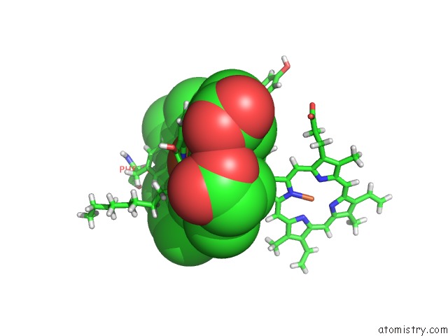
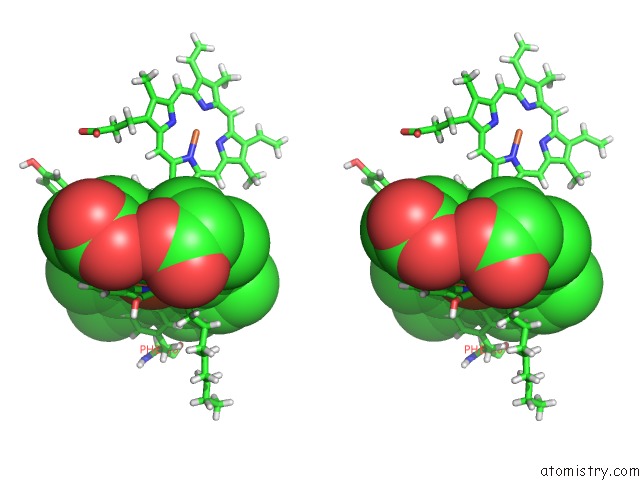
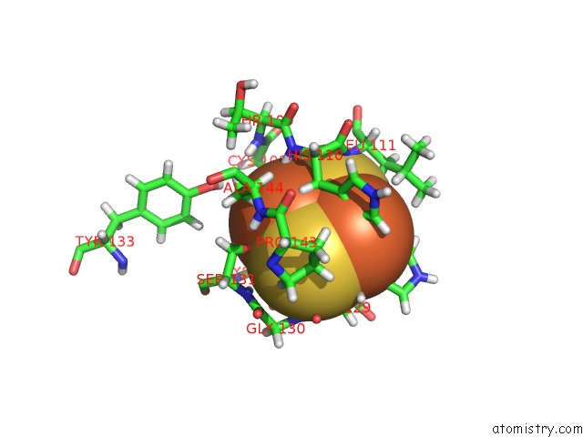
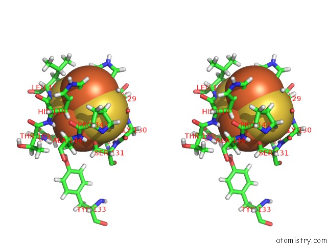
Iron binding site 1 out of 6 in 4h0l
Go back to
Iron binding site 1 out
of 6 in the Cytochrome B6F Complex Crystal Structure From Mastigocladus Laminosus with N-Side Inhibitor Nqno
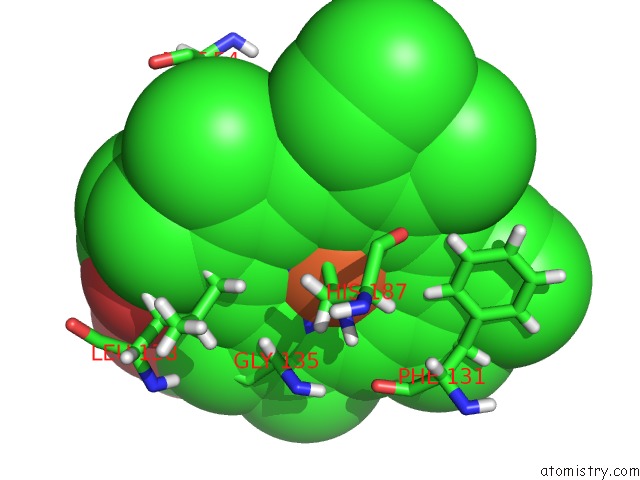
Mono view
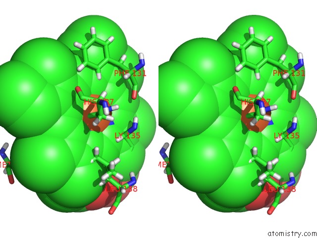
Stereo pair view
Mono view
Stereo pair view
A full contact list of Iron with other atoms in the Fe binding
site number 1 of Cytochrome B6F Complex Crystal Structure From Mastigocladus Laminosus with N-Side Inhibitor Nqno within 5.0Å range:
|
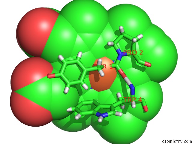
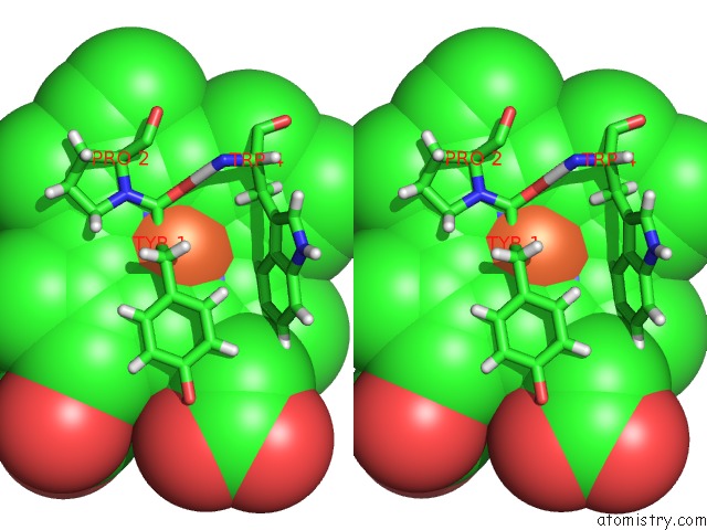
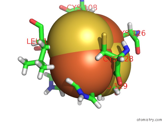
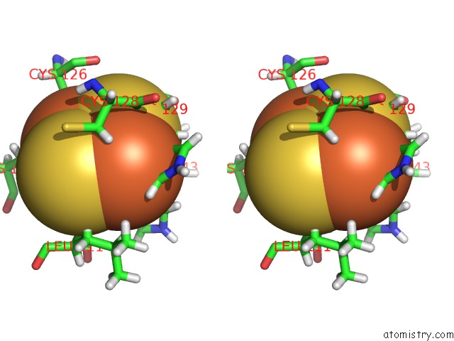
Iron binding site 2 out of 6 in 4h0l
Go back to
Iron binding site 2 out
of 6 in the Cytochrome B6F Complex Crystal Structure From Mastigocladus Laminosus with N-Side Inhibitor Nqno
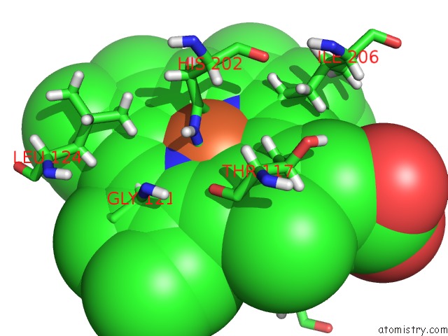
Mono view
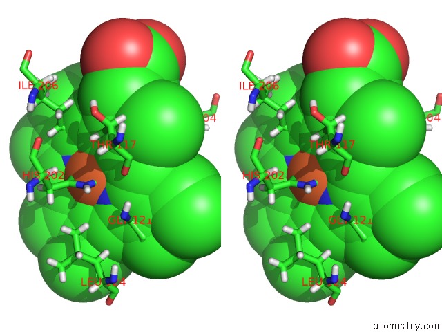
Stereo pair view
Mono view
Stereo pair view
A full contact list of Iron with other atoms in the Fe binding
site number 2 of Cytochrome B6F Complex Crystal Structure From Mastigocladus Laminosus with N-Side Inhibitor Nqno within 5.0Å range:
|
Iron binding site 3 out of 6 in 4h0l
Go back to
Iron binding site 3 out
of 6 in the Cytochrome B6F Complex Crystal Structure From Mastigocladus Laminosus with N-Side Inhibitor Nqno
Mono view
Stereo pair view
Mono view
Stereo pair view
A full contact list of Iron with other atoms in the Fe binding
site number 3 of Cytochrome B6F Complex Crystal Structure From Mastigocladus Laminosus with N-Side Inhibitor Nqno within 5.0Å range:
|
Iron binding site 4 out of 6 in 4h0l
Go back to
Iron binding site 4 out
of 6 in the Cytochrome B6F Complex Crystal Structure From Mastigocladus Laminosus with N-Side Inhibitor Nqno
Mono view
Stereo pair view
Mono view
Stereo pair view
A full contact list of Iron with other atoms in the Fe binding
site number 4 of Cytochrome B6F Complex Crystal Structure From Mastigocladus Laminosus with N-Side Inhibitor Nqno within 5.0Å range:
|
Iron binding site 5 out of 6 in 4h0l
Go back to
Iron binding site 5 out
of 6 in the Cytochrome B6F Complex Crystal Structure From Mastigocladus Laminosus with N-Side Inhibitor Nqno
Mono view
Stereo pair view
Mono view
Stereo pair view
A full contact list of Iron with other atoms in the Fe binding
site number 5 of Cytochrome B6F Complex Crystal Structure From Mastigocladus Laminosus with N-Side Inhibitor Nqno within 5.0Å range:
|
Iron binding site 6 out of 6 in 4h0l
Go back to
Iron binding site 6 out
of 6 in the Cytochrome B6F Complex Crystal Structure From Mastigocladus Laminosus with N-Side Inhibitor Nqno
Mono view
Stereo pair view
Mono view
Stereo pair view
A full contact list of Iron with other atoms in the Fe binding
site number 6 of Cytochrome B6F Complex Crystal Structure From Mastigocladus Laminosus with N-Side Inhibitor Nqno within 5.0Å range:
|
Reference:
S.S.Hasan,
E.Yamashita,
D.Baniulis,
W.A.Cramer.
Quinone-Dependent Proton Transfer Pathways in the Photosynthetic Cytochrome B6F Complex Proc.Natl.Acad.Sci.Usa V. 110 4297 2013.
ISSN: ISSN 0027-8424
PubMed: 23440205
DOI: 10.1073/PNAS.1222248110
Page generated: Mon Aug 5 03:06:59 2024
ISSN: ISSN 0027-8424
PubMed: 23440205
DOI: 10.1073/PNAS.1222248110
Last articles
Zn in 9MJ5Zn in 9HNW
Zn in 9G0L
Zn in 9FNE
Zn in 9DZN
Zn in 9E0I
Zn in 9D32
Zn in 9DAK
Zn in 8ZXC
Zn in 8ZUF