Iron »
PDB 4h9t-4hm6 »
4ha0 »
Iron in PDB 4ha0: Structure of Geobacillus Kaustophilus Lactonase, Mutant R230D with ZN2+
Protein crystallography data
The structure of Structure of Geobacillus Kaustophilus Lactonase, Mutant R230D with ZN2+, PDB code: 4ha0
was solved by
B.Xue,
J.Y.Chow,
W.S.Yew,
R.C.Robinson,
with X-Ray Crystallography technique. A brief refinement statistics is given in the table below:
| Resolution Low / High (Å) | 19.98 / 1.90 |
| Space group | P 2 21 21 |
| Cell size a, b, c (Å), α, β, γ (°) | 69.974, 76.218, 134.765, 90.00, 90.00, 90.00 |
| R / Rfree (%) | 25.9 / 30.3 |
Other elements in 4ha0:
The structure of Structure of Geobacillus Kaustophilus Lactonase, Mutant R230D with ZN2+ also contains other interesting chemical elements:
| Zinc | (Zn) | 2 atoms |
Iron Binding Sites:
The binding sites of Iron atom in the Structure of Geobacillus Kaustophilus Lactonase, Mutant R230D with ZN2+
(pdb code 4ha0). This binding sites where shown within
5.0 Angstroms radius around Iron atom.
In total 2 binding sites of Iron where determined in the Structure of Geobacillus Kaustophilus Lactonase, Mutant R230D with ZN2+, PDB code: 4ha0:
Jump to Iron binding site number: 1; 2;
In total 2 binding sites of Iron where determined in the Structure of Geobacillus Kaustophilus Lactonase, Mutant R230D with ZN2+, PDB code: 4ha0:
Jump to Iron binding site number: 1; 2;
Iron binding site 1 out of 2 in 4ha0
Go back to
Iron binding site 1 out
of 2 in the Structure of Geobacillus Kaustophilus Lactonase, Mutant R230D with ZN2+
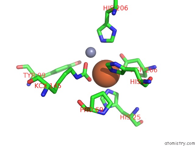
Mono view
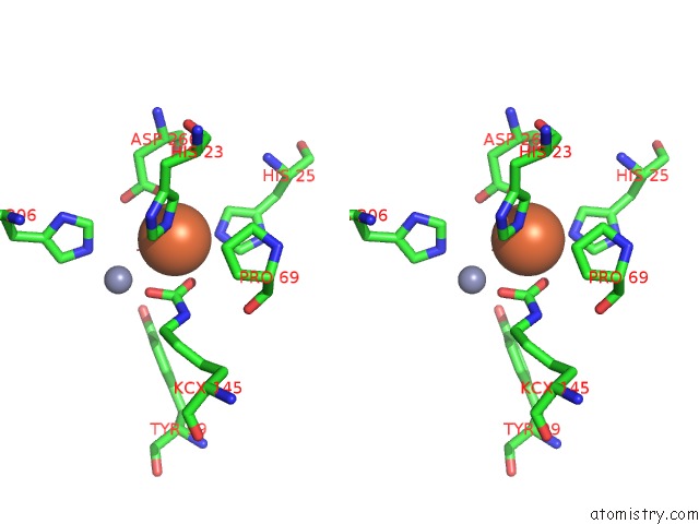
Stereo pair view
Mono view
Stereo pair view
A full contact list of Iron with other atoms in the Fe binding
site number 1 of Structure of Geobacillus Kaustophilus Lactonase, Mutant R230D with ZN2+ within 5.0Å range:
|
Iron binding site 2 out of 2 in 4ha0
Go back to
Iron binding site 2 out
of 2 in the Structure of Geobacillus Kaustophilus Lactonase, Mutant R230D with ZN2+
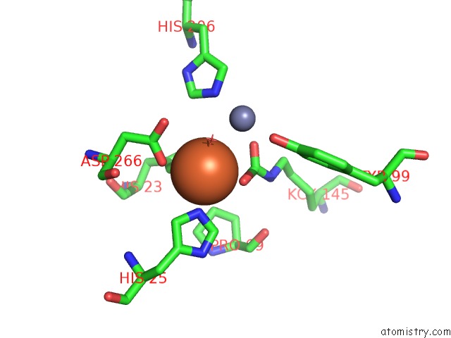
Mono view
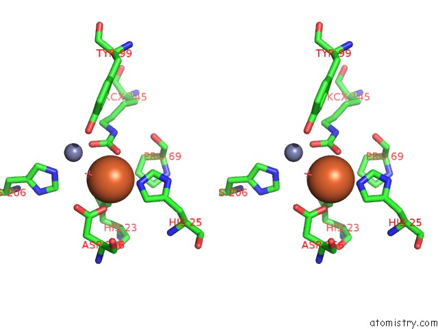
Stereo pair view
Mono view
Stereo pair view
A full contact list of Iron with other atoms in the Fe binding
site number 2 of Structure of Geobacillus Kaustophilus Lactonase, Mutant R230D with ZN2+ within 5.0Å range:
|
Reference:
B.Xue,
J.Y.Chow,
A.Baldansuren,
L.L.Yap,
Y.H.Gan,
S.A.Dikanov,
R.C.Robinson,
W.S.Yew.
Structural Evidence of A Productive Active Site Architecture For An Evolved Quorum-Quenching Gkl Lactonase. Biochemistry V. 52 2359 2013.
ISSN: ISSN 0006-2960
PubMed: 23461395
DOI: 10.1021/BI4000904
Page generated: Mon Aug 5 03:17:15 2024
ISSN: ISSN 0006-2960
PubMed: 23461395
DOI: 10.1021/BI4000904
Last articles
Cl in 7XWRCl in 7XWQ
Cl in 7XWS
Cl in 7XVZ
Cl in 7XVM
Cl in 7XUL
Cl in 7XTQ
Cl in 7XUD
Cl in 7XRB
Cl in 7XUC