Iron »
PDB 4h9t-4hm6 »
4hbi »
Iron in PDB 4hbi: Scapharca Dimeric Hemoglobin, Mutant T72I, Deoxy Form
Protein crystallography data
The structure of Scapharca Dimeric Hemoglobin, Mutant T72I, Deoxy Form, PDB code: 4hbi
was solved by
W.E.Royer Junior,
with X-Ray Crystallography technique. A brief refinement statistics is given in the table below:
| Resolution Low / High (Å) | 10.00 / 1.60 |
| Space group | C 2 2 21 |
| Cell size a, b, c (Å), α, β, γ (°) | 91.760, 44.320, 143.790, 90.00, 90.00, 90.00 |
| R / Rfree (%) | 19.2 / 23.3 |
Iron Binding Sites:
The binding sites of Iron atom in the Scapharca Dimeric Hemoglobin, Mutant T72I, Deoxy Form
(pdb code 4hbi). This binding sites where shown within
5.0 Angstroms radius around Iron atom.
In total 2 binding sites of Iron where determined in the Scapharca Dimeric Hemoglobin, Mutant T72I, Deoxy Form, PDB code: 4hbi:
Jump to Iron binding site number: 1; 2;
In total 2 binding sites of Iron where determined in the Scapharca Dimeric Hemoglobin, Mutant T72I, Deoxy Form, PDB code: 4hbi:
Jump to Iron binding site number: 1; 2;
Iron binding site 1 out of 2 in 4hbi
Go back to
Iron binding site 1 out
of 2 in the Scapharca Dimeric Hemoglobin, Mutant T72I, Deoxy Form
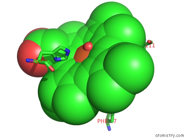
Mono view
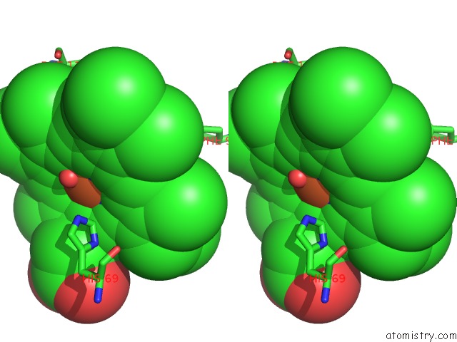
Stereo pair view
Mono view
Stereo pair view
A full contact list of Iron with other atoms in the Fe binding
site number 1 of Scapharca Dimeric Hemoglobin, Mutant T72I, Deoxy Form within 5.0Å range:
|
Iron binding site 2 out of 2 in 4hbi
Go back to
Iron binding site 2 out
of 2 in the Scapharca Dimeric Hemoglobin, Mutant T72I, Deoxy Form
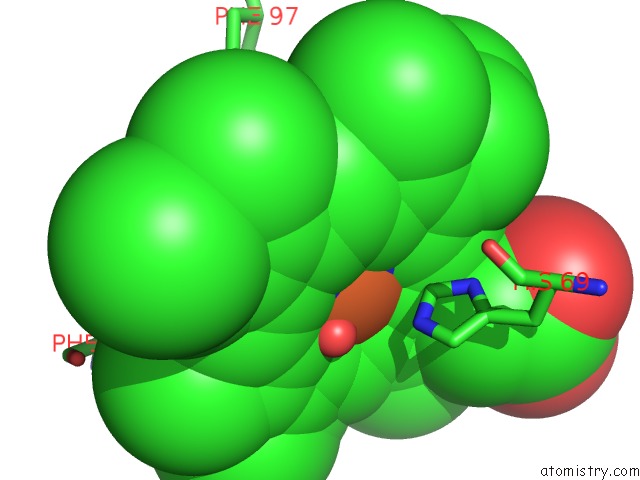
Mono view
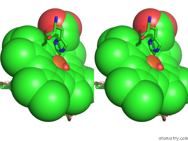
Stereo pair view
Mono view
Stereo pair view
A full contact list of Iron with other atoms in the Fe binding
site number 2 of Scapharca Dimeric Hemoglobin, Mutant T72I, Deoxy Form within 5.0Å range:
|
Reference:
A.Pardanani,
A.Gambacurta,
F.Ascoli,
W.E.Royer Jr..
Mutational Destabilization of the Critical Interface Water Cluster in Scapharca Dimeric Hemoglobin: Structural Basis For Altered Allosteric Activity. J.Mol.Biol. V. 284 729 1998.
ISSN: ISSN 0022-2836
PubMed: 9826511
DOI: 10.1006/JMBI.1998.2195
Page generated: Mon Aug 5 03:19:24 2024
ISSN: ISSN 0022-2836
PubMed: 9826511
DOI: 10.1006/JMBI.1998.2195
Last articles
Zn in 9J0NZn in 9J0O
Zn in 9J0P
Zn in 9FJX
Zn in 9EKB
Zn in 9C0F
Zn in 9CAH
Zn in 9CH0
Zn in 9CH3
Zn in 9CH1