Iron »
PDB 4h9t-4hm6 »
4hgh »
Iron in PDB 4hgh: Crystal Structure of P450 BM3 5F5 Heme Domain Variant Complexed with Styrene (Dataset I)
Enzymatic activity of Crystal Structure of P450 BM3 5F5 Heme Domain Variant Complexed with Styrene (Dataset I)
All present enzymatic activity of Crystal Structure of P450 BM3 5F5 Heme Domain Variant Complexed with Styrene (Dataset I):
1.14.14.1; 1.6.2.4;
1.14.14.1; 1.6.2.4;
Protein crystallography data
The structure of Crystal Structure of P450 BM3 5F5 Heme Domain Variant Complexed with Styrene (Dataset I), PDB code: 4hgh
was solved by
A.Shehzad,
S.Panneerselvam,
M.Bocola,
J.Mueller-Dieckmann,
M.Wilmanns,
U.Schwaneberg,
with X-Ray Crystallography technique. A brief refinement statistics is given in the table below:
| Resolution Low / High (Å) | 19.87 / 1.40 |
| Space group | P 1 21 1 |
| Cell size a, b, c (Å), α, β, γ (°) | 59.090, 149.270, 65.160, 90.00, 98.38, 90.00 |
| R / Rfree (%) | 19 / 20.4 |
Iron Binding Sites:
The binding sites of Iron atom in the Crystal Structure of P450 BM3 5F5 Heme Domain Variant Complexed with Styrene (Dataset I)
(pdb code 4hgh). This binding sites where shown within
5.0 Angstroms radius around Iron atom.
In total 2 binding sites of Iron where determined in the Crystal Structure of P450 BM3 5F5 Heme Domain Variant Complexed with Styrene (Dataset I), PDB code: 4hgh:
Jump to Iron binding site number: 1; 2;
In total 2 binding sites of Iron where determined in the Crystal Structure of P450 BM3 5F5 Heme Domain Variant Complexed with Styrene (Dataset I), PDB code: 4hgh:
Jump to Iron binding site number: 1; 2;
Iron binding site 1 out of 2 in 4hgh
Go back to
Iron binding site 1 out
of 2 in the Crystal Structure of P450 BM3 5F5 Heme Domain Variant Complexed with Styrene (Dataset I)
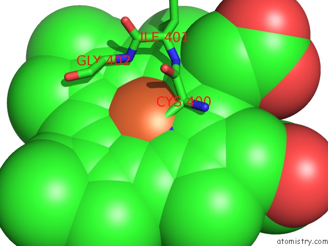
Mono view
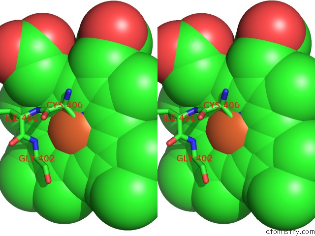
Stereo pair view
Mono view
Stereo pair view
A full contact list of Iron with other atoms in the Fe binding
site number 1 of Crystal Structure of P450 BM3 5F5 Heme Domain Variant Complexed with Styrene (Dataset I) within 5.0Å range:
|
Iron binding site 2 out of 2 in 4hgh
Go back to
Iron binding site 2 out
of 2 in the Crystal Structure of P450 BM3 5F5 Heme Domain Variant Complexed with Styrene (Dataset I)
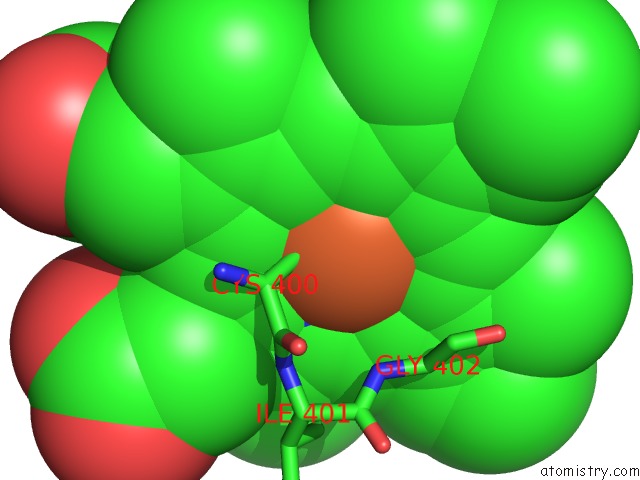
Mono view
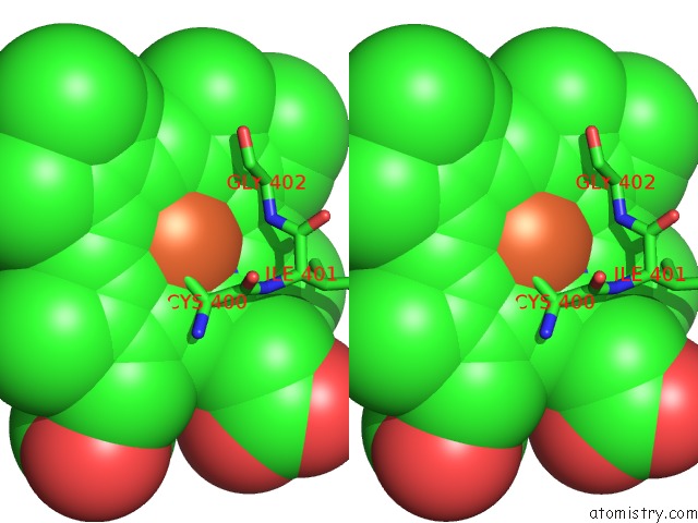
Stereo pair view
Mono view
Stereo pair view
A full contact list of Iron with other atoms in the Fe binding
site number 2 of Crystal Structure of P450 BM3 5F5 Heme Domain Variant Complexed with Styrene (Dataset I) within 5.0Å range:
|
Reference:
A.Shehzad,
S.Panneerselvam,
M.Linow,
M.Bocola,
D.Roccatano,
J.Mueller-Dieckmann,
M.Wilmanns,
U.Schwaneberg.
P450 BM3 Crystal Structures Reveal the Role of the Charged Surface Residue Lys/ARG184 in Inversion of Enantioselective Styrene Epoxidation. Chem.Commun.(Camb.) V. 49 4694 2013.
ISSN: ISSN 1359-7345
PubMed: 23589805
DOI: 10.1039/C3CC39076D
Page generated: Mon Aug 5 03:20:53 2024
ISSN: ISSN 1359-7345
PubMed: 23589805
DOI: 10.1039/C3CC39076D
Last articles
Zn in 9J0NZn in 9J0O
Zn in 9J0P
Zn in 9FJX
Zn in 9EKB
Zn in 9C0F
Zn in 9CAH
Zn in 9CH0
Zn in 9CH3
Zn in 9CH1