Iron »
PDB 4hm7-4ies »
4i4h »
Iron in PDB 4i4h: Crystal Structure of CYP3A4 Ligated to Pyridine-Substituted Desoxyritonavir
Enzymatic activity of Crystal Structure of CYP3A4 Ligated to Pyridine-Substituted Desoxyritonavir
All present enzymatic activity of Crystal Structure of CYP3A4 Ligated to Pyridine-Substituted Desoxyritonavir:
1.14.13.157; 1.14.13.32; 1.14.13.67; 1.14.13.97;
1.14.13.157; 1.14.13.32; 1.14.13.67; 1.14.13.97;
Protein crystallography data
The structure of Crystal Structure of CYP3A4 Ligated to Pyridine-Substituted Desoxyritonavir, PDB code: 4i4h
was solved by
I.F.Sevrioukova,
T.L.Poulos,
with X-Ray Crystallography technique. A brief refinement statistics is given in the table below:
| Resolution Low / High (Å) | 65.70 / 2.90 |
| Space group | I 2 2 2 |
| Cell size a, b, c (Å), α, β, γ (°) | 77.760, 100.060, 131.400, 90.00, 90.00, 90.00 |
| R / Rfree (%) | 19.5 / 26.9 |
Iron Binding Sites:
The binding sites of Iron atom in the Crystal Structure of CYP3A4 Ligated to Pyridine-Substituted Desoxyritonavir
(pdb code 4i4h). This binding sites where shown within
5.0 Angstroms radius around Iron atom.
In total only one binding site of Iron was determined in the Crystal Structure of CYP3A4 Ligated to Pyridine-Substituted Desoxyritonavir, PDB code: 4i4h:
In total only one binding site of Iron was determined in the Crystal Structure of CYP3A4 Ligated to Pyridine-Substituted Desoxyritonavir, PDB code: 4i4h:
Iron binding site 1 out of 1 in 4i4h
Go back to
Iron binding site 1 out
of 1 in the Crystal Structure of CYP3A4 Ligated to Pyridine-Substituted Desoxyritonavir

Mono view
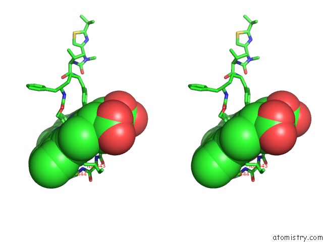
Stereo pair view
Mono view
Stereo pair view
A full contact list of Iron with other atoms in the Fe binding
site number 1 of Crystal Structure of CYP3A4 Ligated to Pyridine-Substituted Desoxyritonavir within 5.0Å range:
|
Reference:
I.F.Sevrioukova,
T.L.Poulos.
Pyridine-Substituted Desoxyritonavir Is A More Potent Inhibitor of Cytochrome P450 3A4 Than Ritonavir. J.Med.Chem. V. 56 3733 2013.
ISSN: ISSN 0022-2623
PubMed: 23586711
DOI: 10.1021/JM400288Z
Page generated: Mon Aug 5 03:53:06 2024
ISSN: ISSN 0022-2623
PubMed: 23586711
DOI: 10.1021/JM400288Z
Last articles
Zn in 9J0NZn in 9J0O
Zn in 9J0P
Zn in 9FJX
Zn in 9EKB
Zn in 9C0F
Zn in 9CAH
Zn in 9CH0
Zn in 9CH3
Zn in 9CH1