Iron »
PDB 4iet-4ixq »
4ixk »
Iron in PDB 4ixk: Anaerobic Crystal Structure of Iron Soaked (2 H) Ferritin From Pseudo- Nitzschia Multiseries
Protein crystallography data
The structure of Anaerobic Crystal Structure of Iron Soaked (2 H) Ferritin From Pseudo- Nitzschia Multiseries, PDB code: 4ixk
was solved by
S.Pfaffen,
M.E.P.Murphy,
with X-Ray Crystallography technique. A brief refinement statistics is given in the table below:
| Resolution Low / High (Å) | 48.40 / 2.10 |
| Space group | P 2 3 |
| Cell size a, b, c (Å), α, β, γ (°) | 174.323, 174.323, 174.323, 90.00, 90.00, 90.00 |
| R / Rfree (%) | 20.1 / 24.7 |
Iron Binding Sites:
The binding sites of Iron atom in the Anaerobic Crystal Structure of Iron Soaked (2 H) Ferritin From Pseudo- Nitzschia Multiseries
(pdb code 4ixk). This binding sites where shown within
5.0 Angstroms radius around Iron atom.
In total 8 binding sites of Iron where determined in the Anaerobic Crystal Structure of Iron Soaked (2 H) Ferritin From Pseudo- Nitzschia Multiseries, PDB code: 4ixk:
Jump to Iron binding site number: 1; 2; 3; 4; 5; 6; 7; 8;
In total 8 binding sites of Iron where determined in the Anaerobic Crystal Structure of Iron Soaked (2 H) Ferritin From Pseudo- Nitzschia Multiseries, PDB code: 4ixk:
Jump to Iron binding site number: 1; 2; 3; 4; 5; 6; 7; 8;
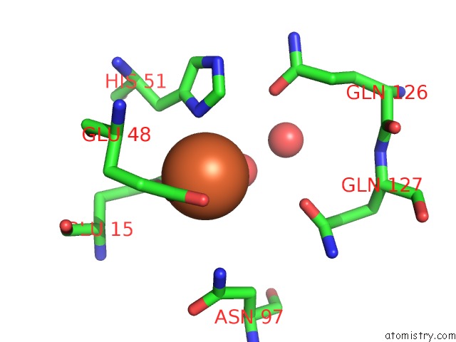
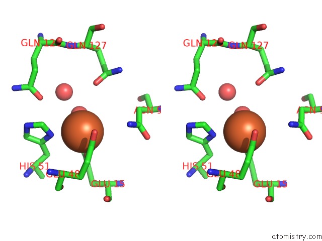
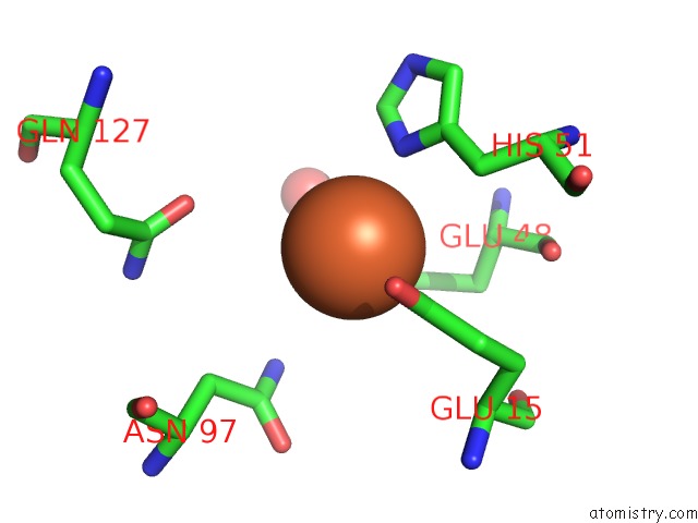
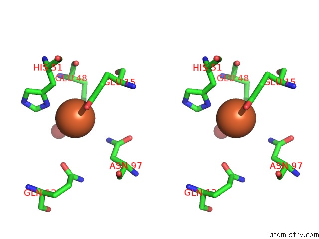
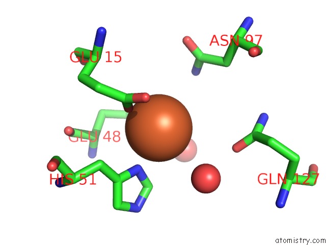
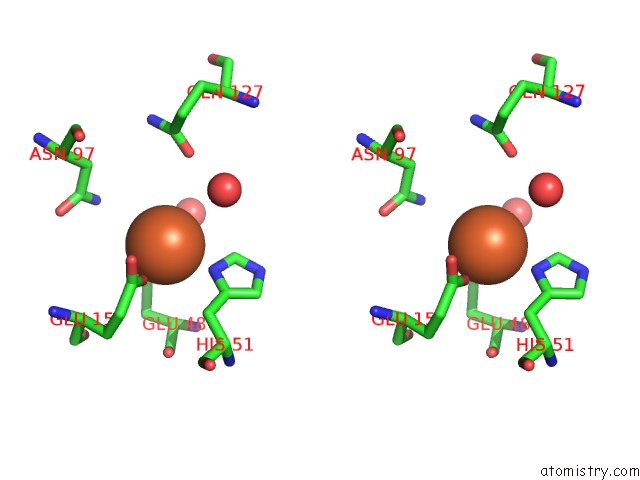
Iron binding site 1 out of 8 in 4ixk
Go back to
Iron binding site 1 out
of 8 in the Anaerobic Crystal Structure of Iron Soaked (2 H) Ferritin From Pseudo- Nitzschia Multiseries
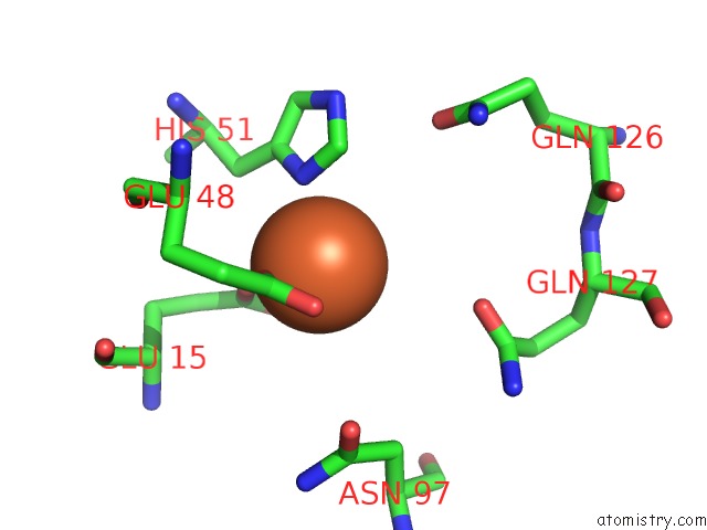
Mono view
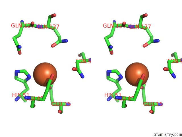
Stereo pair view
Mono view
Stereo pair view
A full contact list of Iron with other atoms in the Fe binding
site number 1 of Anaerobic Crystal Structure of Iron Soaked (2 H) Ferritin From Pseudo- Nitzschia Multiseries within 5.0Å range:
|
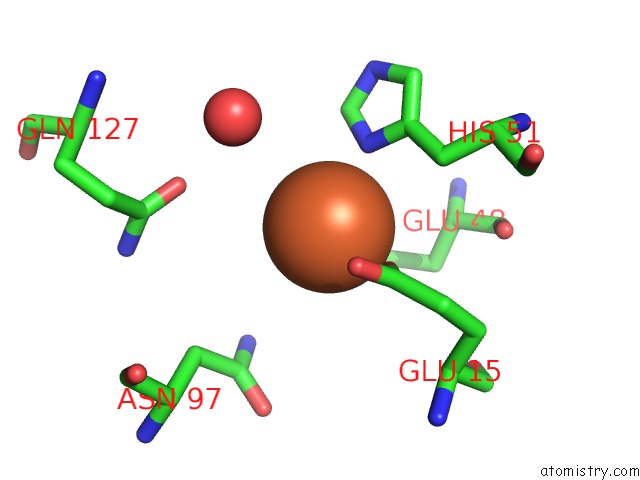
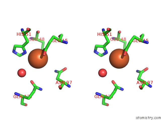
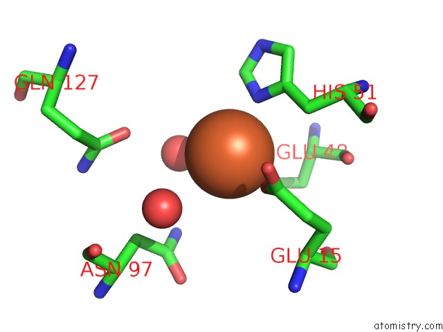
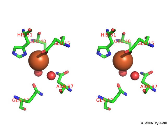
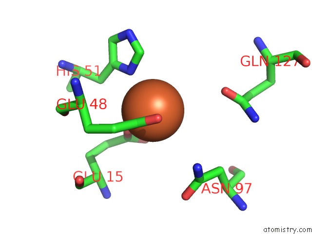
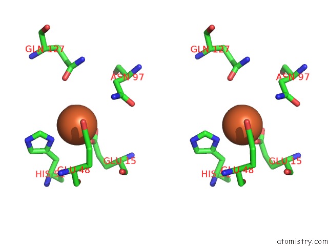
Iron binding site 2 out of 8 in 4ixk
Go back to
Iron binding site 2 out
of 8 in the Anaerobic Crystal Structure of Iron Soaked (2 H) Ferritin From Pseudo- Nitzschia Multiseries
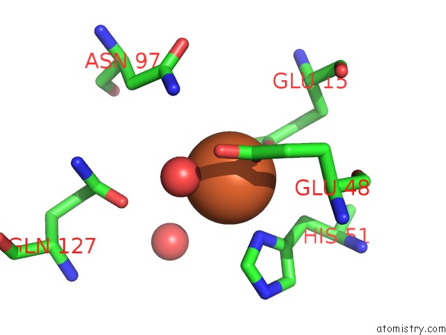
Mono view
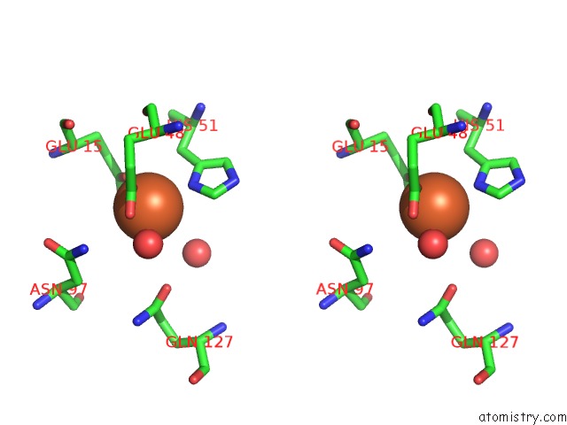
Stereo pair view
Mono view
Stereo pair view
A full contact list of Iron with other atoms in the Fe binding
site number 2 of Anaerobic Crystal Structure of Iron Soaked (2 H) Ferritin From Pseudo- Nitzschia Multiseries within 5.0Å range:
|
Iron binding site 3 out of 8 in 4ixk
Go back to
Iron binding site 3 out
of 8 in the Anaerobic Crystal Structure of Iron Soaked (2 H) Ferritin From Pseudo- Nitzschia Multiseries
Mono view
Stereo pair view
Mono view
Stereo pair view
A full contact list of Iron with other atoms in the Fe binding
site number 3 of Anaerobic Crystal Structure of Iron Soaked (2 H) Ferritin From Pseudo- Nitzschia Multiseries within 5.0Å range:
|
Iron binding site 4 out of 8 in 4ixk
Go back to
Iron binding site 4 out
of 8 in the Anaerobic Crystal Structure of Iron Soaked (2 H) Ferritin From Pseudo- Nitzschia Multiseries
Mono view
Stereo pair view
Mono view
Stereo pair view
A full contact list of Iron with other atoms in the Fe binding
site number 4 of Anaerobic Crystal Structure of Iron Soaked (2 H) Ferritin From Pseudo- Nitzschia Multiseries within 5.0Å range:
|
Iron binding site 5 out of 8 in 4ixk
Go back to
Iron binding site 5 out
of 8 in the Anaerobic Crystal Structure of Iron Soaked (2 H) Ferritin From Pseudo- Nitzschia Multiseries
Mono view
Stereo pair view
Mono view
Stereo pair view
A full contact list of Iron with other atoms in the Fe binding
site number 5 of Anaerobic Crystal Structure of Iron Soaked (2 H) Ferritin From Pseudo- Nitzschia Multiseries within 5.0Å range:
|
Iron binding site 6 out of 8 in 4ixk
Go back to
Iron binding site 6 out
of 8 in the Anaerobic Crystal Structure of Iron Soaked (2 H) Ferritin From Pseudo- Nitzschia Multiseries
Mono view
Stereo pair view
Mono view
Stereo pair view
A full contact list of Iron with other atoms in the Fe binding
site number 6 of Anaerobic Crystal Structure of Iron Soaked (2 H) Ferritin From Pseudo- Nitzschia Multiseries within 5.0Å range:
|
Iron binding site 7 out of 8 in 4ixk
Go back to
Iron binding site 7 out
of 8 in the Anaerobic Crystal Structure of Iron Soaked (2 H) Ferritin From Pseudo- Nitzschia Multiseries
Mono view
Stereo pair view
Mono view
Stereo pair view
A full contact list of Iron with other atoms in the Fe binding
site number 7 of Anaerobic Crystal Structure of Iron Soaked (2 H) Ferritin From Pseudo- Nitzschia Multiseries within 5.0Å range:
|
Iron binding site 8 out of 8 in 4ixk
Go back to
Iron binding site 8 out
of 8 in the Anaerobic Crystal Structure of Iron Soaked (2 H) Ferritin From Pseudo- Nitzschia Multiseries
Mono view
Stereo pair view
Mono view
Stereo pair view
A full contact list of Iron with other atoms in the Fe binding
site number 8 of Anaerobic Crystal Structure of Iron Soaked (2 H) Ferritin From Pseudo- Nitzschia Multiseries within 5.0Å range:
|
Reference:
S.Pfaffen,
R.Abdulqadir,
N.E.Le Brun,
M.E.Murphy.
Mechanism of Ferrous Iron Binding and Oxidation By Ferritin From A Pennate Diatom. J.Biol.Chem. V. 288 14917 2013.
ISSN: ISSN 0021-9258
PubMed: 23548912
DOI: 10.1074/JBC.M113.454496
Page generated: Mon Aug 5 04:18:02 2024
ISSN: ISSN 0021-9258
PubMed: 23548912
DOI: 10.1074/JBC.M113.454496
Last articles
Zn in 9J0NZn in 9J0O
Zn in 9J0P
Zn in 9FJX
Zn in 9EKB
Zn in 9C0F
Zn in 9CAH
Zn in 9CH0
Zn in 9CH3
Zn in 9CH1