Iron »
PDB 4ixr-4jn0 »
4jm8 »
Iron in PDB 4jm8: Crystal Structure of Cytochrome C Peroxidase W191G-Gateless in Complex with 2,6-Diaminopyridine
Enzymatic activity of Crystal Structure of Cytochrome C Peroxidase W191G-Gateless in Complex with 2,6-Diaminopyridine
All present enzymatic activity of Crystal Structure of Cytochrome C Peroxidase W191G-Gateless in Complex with 2,6-Diaminopyridine:
1.11.1.5;
1.11.1.5;
Protein crystallography data
The structure of Crystal Structure of Cytochrome C Peroxidase W191G-Gateless in Complex with 2,6-Diaminopyridine, PDB code: 4jm8
was solved by
S.E.Boyce,
M.Fischer,
I.Fish,
with X-Ray Crystallography technique. A brief refinement statistics is given in the table below:
| Resolution Low / High (Å) | 30.05 / 1.30 |
| Space group | P 21 21 21 |
| Cell size a, b, c (Å), α, β, γ (°) | 50.843, 74.519, 106.464, 90.00, 90.00, 90.00 |
| R / Rfree (%) | 11.7 / 13.7 |
Iron Binding Sites:
The binding sites of Iron atom in the Crystal Structure of Cytochrome C Peroxidase W191G-Gateless in Complex with 2,6-Diaminopyridine
(pdb code 4jm8). This binding sites where shown within
5.0 Angstroms radius around Iron atom.
In total only one binding site of Iron was determined in the Crystal Structure of Cytochrome C Peroxidase W191G-Gateless in Complex with 2,6-Diaminopyridine, PDB code: 4jm8:
In total only one binding site of Iron was determined in the Crystal Structure of Cytochrome C Peroxidase W191G-Gateless in Complex with 2,6-Diaminopyridine, PDB code: 4jm8:
Iron binding site 1 out of 1 in 4jm8
Go back to
Iron binding site 1 out
of 1 in the Crystal Structure of Cytochrome C Peroxidase W191G-Gateless in Complex with 2,6-Diaminopyridine
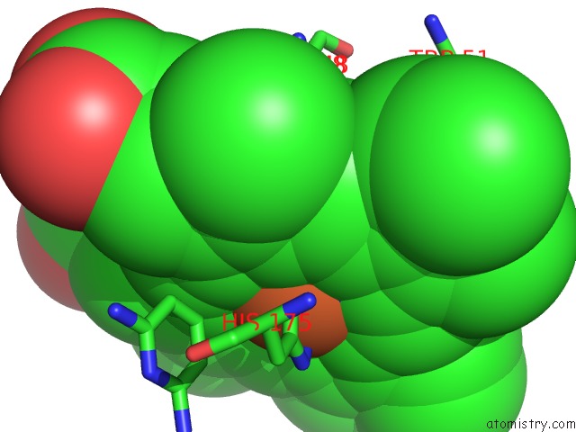
Mono view
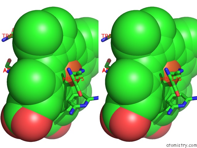
Stereo pair view
Mono view
Stereo pair view
A full contact list of Iron with other atoms in the Fe binding
site number 1 of Crystal Structure of Cytochrome C Peroxidase W191G-Gateless in Complex with 2,6-Diaminopyridine within 5.0Å range:
|
Reference:
G.J.Rocklin,
S.E.Boyce,
M.Fischer,
I.Fish,
D.L.Mobley,
B.K.Shoichet,
K.A.Dill.
Blind Prediction of Charged Ligand Binding Affinities in A Model Binding Site. J.Mol.Biol. V. 425 4569 2013.
ISSN: ISSN 0022-2836
PubMed: 23896298
DOI: 10.1016/J.JMB.2013.07.030
Page generated: Mon Aug 5 04:43:32 2024
ISSN: ISSN 0022-2836
PubMed: 23896298
DOI: 10.1016/J.JMB.2013.07.030
Last articles
Zn in 9J0NZn in 9J0O
Zn in 9J0P
Zn in 9FJX
Zn in 9EKB
Zn in 9C0F
Zn in 9CAH
Zn in 9CH0
Zn in 9CH3
Zn in 9CH1