Iron »
PDB 4ixr-4jn0 »
4jmw »
Iron in PDB 4jmw: Crystal Structure of Cytochrome C Peroxidase W191G-Gateless in Complex with Phenol
Enzymatic activity of Crystal Structure of Cytochrome C Peroxidase W191G-Gateless in Complex with Phenol
All present enzymatic activity of Crystal Structure of Cytochrome C Peroxidase W191G-Gateless in Complex with Phenol:
1.11.1.5;
1.11.1.5;
Protein crystallography data
The structure of Crystal Structure of Cytochrome C Peroxidase W191G-Gateless in Complex with Phenol, PDB code: 4jmw
was solved by
M.Fischer,
I.Fish,
with X-Ray Crystallography technique. A brief refinement statistics is given in the table below:
| Resolution Low / High (Å) | 32.06 / 1.19 |
| Space group | P 21 21 21 |
| Cell size a, b, c (Å), α, β, γ (°) | 51.050, 74.410, 106.600, 90.00, 90.00, 90.00 |
| R / Rfree (%) | 12.5 / 15.1 |
Iron Binding Sites:
The binding sites of Iron atom in the Crystal Structure of Cytochrome C Peroxidase W191G-Gateless in Complex with Phenol
(pdb code 4jmw). This binding sites where shown within
5.0 Angstroms radius around Iron atom.
In total only one binding site of Iron was determined in the Crystal Structure of Cytochrome C Peroxidase W191G-Gateless in Complex with Phenol, PDB code: 4jmw:
In total only one binding site of Iron was determined in the Crystal Structure of Cytochrome C Peroxidase W191G-Gateless in Complex with Phenol, PDB code: 4jmw:
Iron binding site 1 out of 1 in 4jmw
Go back to
Iron binding site 1 out
of 1 in the Crystal Structure of Cytochrome C Peroxidase W191G-Gateless in Complex with Phenol
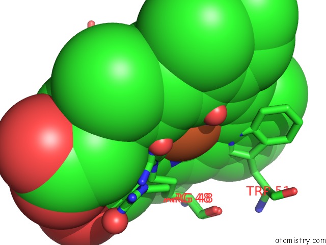
Mono view
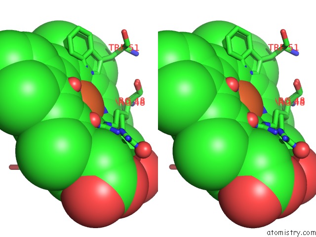
Stereo pair view
Mono view
Stereo pair view
A full contact list of Iron with other atoms in the Fe binding
site number 1 of Crystal Structure of Cytochrome C Peroxidase W191G-Gateless in Complex with Phenol within 5.0Å range:
|
Reference:
G.J.Rocklin,
S.E.Boyce,
M.Fischer,
I.Fish,
D.L.Mobley,
B.K.Shoichet,
K.A.Dill.
Blind Prediction of Charged Ligand Binding Affinities in A Model Binding Site. J.Mol.Biol. V. 425 4569 2013.
ISSN: ISSN 0022-2836
PubMed: 23896298
DOI: 10.1016/J.JMB.2013.07.030
Page generated: Mon Aug 5 04:45:17 2024
ISSN: ISSN 0022-2836
PubMed: 23896298
DOI: 10.1016/J.JMB.2013.07.030
Last articles
Cl in 5SDKCl in 5SDI
Cl in 5SDJ
Cl in 5SDH
Cl in 5SDD
Cl in 5SDE
Cl in 5SDF
Cl in 5SDG
Cl in 5SDC
Cl in 5SD6