Iron »
PDB 4jo0-4k5d »
4jy9 »
Iron in PDB 4jy9: X-Ray Snapshots of Possible Intermediates in the Time Course of Synthesis and Degradation of Protein-Bound FE4S4 Clusters
Protein crystallography data
The structure of X-Ray Snapshots of Possible Intermediates in the Time Course of Synthesis and Degradation of Protein-Bound FE4S4 Clusters, PDB code: 4jy9
was solved by
Y.Nicolet,
R.Rohac,
L.Martin,
J.C.Fontecilla-Camps,
with X-Ray Crystallography technique. A brief refinement statistics is given in the table below:
| Resolution Low / High (Å) | 58.22 / 1.60 |
| Space group | P 21 21 21 |
| Cell size a, b, c (Å), α, β, γ (°) | 51.247, 78.950, 86.145, 90.00, 90.00, 90.00 |
| R / Rfree (%) | 10.2 / 15.6 |
Other elements in 4jy9:
The structure of X-Ray Snapshots of Possible Intermediates in the Time Course of Synthesis and Degradation of Protein-Bound FE4S4 Clusters also contains other interesting chemical elements:
| Chlorine | (Cl) | 4 atoms |
Iron Binding Sites:
The binding sites of Iron atom in the X-Ray Snapshots of Possible Intermediates in the Time Course of Synthesis and Degradation of Protein-Bound FE4S4 Clusters
(pdb code 4jy9). This binding sites where shown within
5.0 Angstroms radius around Iron atom.
In total 6 binding sites of Iron where determined in the X-Ray Snapshots of Possible Intermediates in the Time Course of Synthesis and Degradation of Protein-Bound FE4S4 Clusters, PDB code: 4jy9:
Jump to Iron binding site number: 1; 2; 3; 4; 5; 6;
In total 6 binding sites of Iron where determined in the X-Ray Snapshots of Possible Intermediates in the Time Course of Synthesis and Degradation of Protein-Bound FE4S4 Clusters, PDB code: 4jy9:
Jump to Iron binding site number: 1; 2; 3; 4; 5; 6;
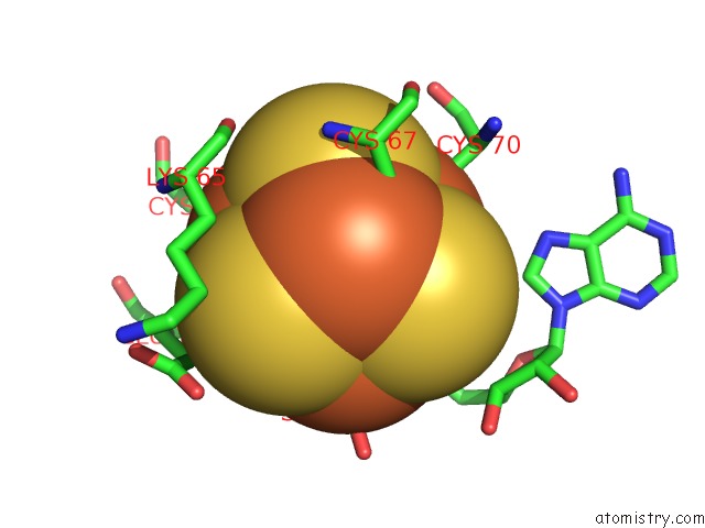
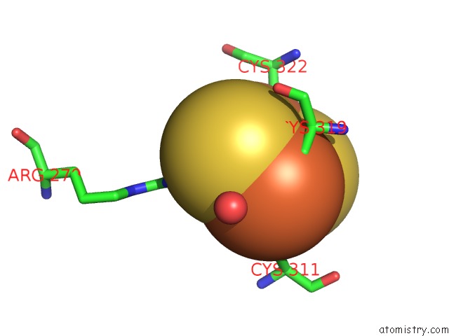
Iron binding site 1 out of 6 in 4jy9
Go back to
Iron binding site 1 out
of 6 in the X-Ray Snapshots of Possible Intermediates in the Time Course of Synthesis and Degradation of Protein-Bound FE4S4 Clusters
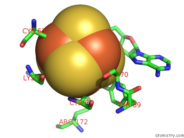
Mono view
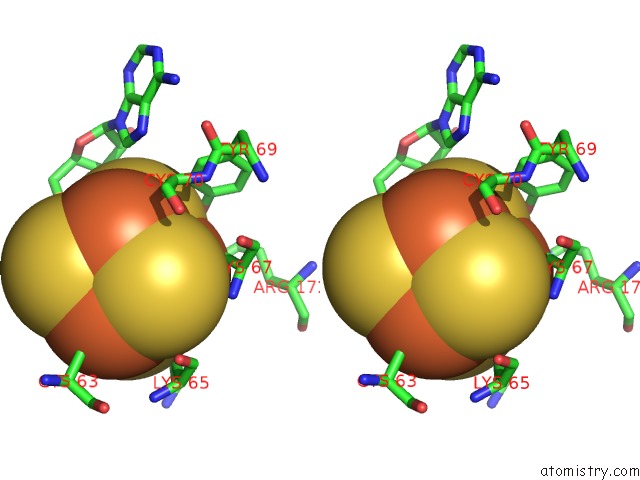
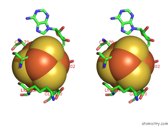
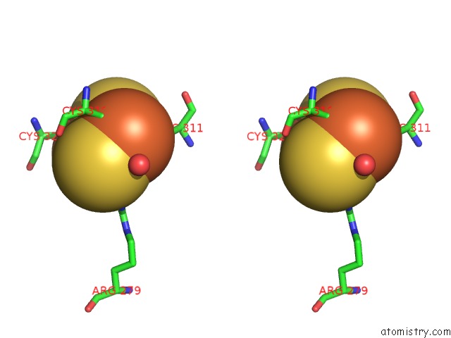
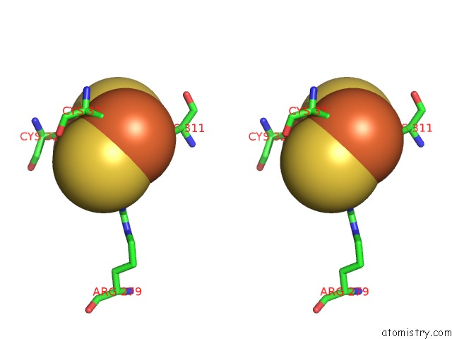
Stereo pair view
Mono view
Stereo pair view
A full contact list of Iron with other atoms in the Fe binding
site number 1 of X-Ray Snapshots of Possible Intermediates in the Time Course of Synthesis and Degradation of Protein-Bound FE4S4 Clusters within 5.0Å range:
|
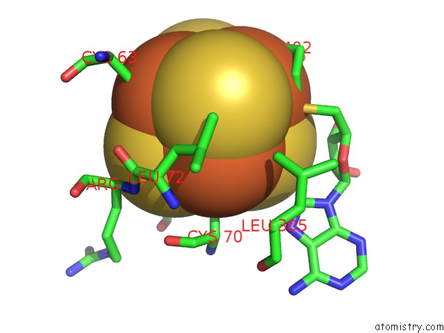
Iron binding site 2 out of 6 in 4jy9
Go back to
Iron binding site 2 out
of 6 in the X-Ray Snapshots of Possible Intermediates in the Time Course of Synthesis and Degradation of Protein-Bound FE4S4 Clusters
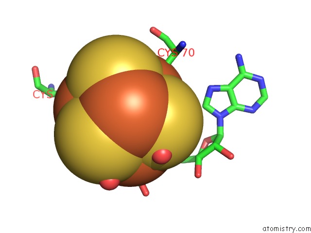
Mono view
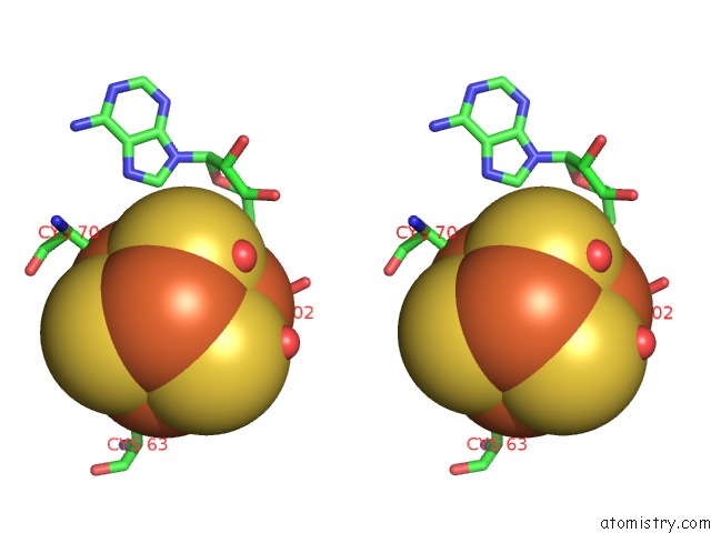
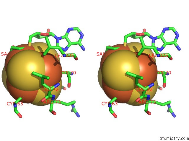
Stereo pair view
Mono view
Stereo pair view
A full contact list of Iron with other atoms in the Fe binding
site number 2 of X-Ray Snapshots of Possible Intermediates in the Time Course of Synthesis and Degradation of Protein-Bound FE4S4 Clusters within 5.0Å range:
|
Iron binding site 3 out of 6 in 4jy9
Go back to
Iron binding site 3 out
of 6 in the X-Ray Snapshots of Possible Intermediates in the Time Course of Synthesis and Degradation of Protein-Bound FE4S4 Clusters
Mono view
Stereo pair view
Mono view
Stereo pair view
A full contact list of Iron with other atoms in the Fe binding
site number 3 of X-Ray Snapshots of Possible Intermediates in the Time Course of Synthesis and Degradation of Protein-Bound FE4S4 Clusters within 5.0Å range:
|
Iron binding site 4 out of 6 in 4jy9
Go back to
Iron binding site 4 out
of 6 in the X-Ray Snapshots of Possible Intermediates in the Time Course of Synthesis and Degradation of Protein-Bound FE4S4 Clusters
Mono view
Stereo pair view
Mono view
Stereo pair view
A full contact list of Iron with other atoms in the Fe binding
site number 4 of X-Ray Snapshots of Possible Intermediates in the Time Course of Synthesis and Degradation of Protein-Bound FE4S4 Clusters within 5.0Å range:
|
Iron binding site 5 out of 6 in 4jy9
Go back to
Iron binding site 5 out
of 6 in the X-Ray Snapshots of Possible Intermediates in the Time Course of Synthesis and Degradation of Protein-Bound FE4S4 Clusters
Mono view
Stereo pair view
Mono view
Stereo pair view
A full contact list of Iron with other atoms in the Fe binding
site number 5 of X-Ray Snapshots of Possible Intermediates in the Time Course of Synthesis and Degradation of Protein-Bound FE4S4 Clusters within 5.0Å range:
|
Iron binding site 6 out of 6 in 4jy9
Go back to
Iron binding site 6 out
of 6 in the X-Ray Snapshots of Possible Intermediates in the Time Course of Synthesis and Degradation of Protein-Bound FE4S4 Clusters

Mono view
Stereo pair view
Mono view
Stereo pair view
A full contact list of Iron with other atoms in the Fe binding
site number 6 of X-Ray Snapshots of Possible Intermediates in the Time Course of Synthesis and Degradation of Protein-Bound FE4S4 Clusters within 5.0Å range:
|
Reference:
Y.Nicolet,
R.Rohac,
L.Martin,
J.C.Fontecilla-Camps.
X-Ray Snapshots of Possible Intermediates in the Time Course of Synthesis and Degradation of Protein-Bound FE4S4 Clusters. Proc.Natl.Acad.Sci.Usa V. 110 7188 2013.
ISSN: ISSN 0027-8424
PubMed: 23596207
DOI: 10.1073/PNAS.1302388110
Page generated: Mon Aug 5 04:53:35 2024
ISSN: ISSN 0027-8424
PubMed: 23596207
DOI: 10.1073/PNAS.1302388110
Last articles
Zn in 9MJ5Zn in 9HNW
Zn in 9G0L
Zn in 9FNE
Zn in 9DZN
Zn in 9E0I
Zn in 9D32
Zn in 9DAK
Zn in 8ZXC
Zn in 8ZUF