Iron »
PDB 4jo0-4k5d »
4k3i »
Iron in PDB 4k3i: Crystal Structure of the Quinol Form of Methylamine Dehydrogenase in Complex with the Diferrous Form of Maug, C2 Space Group
Enzymatic activity of Crystal Structure of the Quinol Form of Methylamine Dehydrogenase in Complex with the Diferrous Form of Maug, C2 Space Group
All present enzymatic activity of Crystal Structure of the Quinol Form of Methylamine Dehydrogenase in Complex with the Diferrous Form of Maug, C2 Space Group:
1.4.9.1; 1.4.99.3;
1.4.9.1; 1.4.99.3;
Protein crystallography data
The structure of Crystal Structure of the Quinol Form of Methylamine Dehydrogenase in Complex with the Diferrous Form of Maug, C2 Space Group, PDB code: 4k3i
was solved by
E.Y.Yukl,
C.M.Wilmot,
with X-Ray Crystallography technique. A brief refinement statistics is given in the table below:
| Resolution Low / High (Å) | 43.06 / 2.00 |
| Space group | C 1 2 1 |
| Cell size a, b, c (Å), α, β, γ (°) | 346.356, 55.558, 112.548, 90.00, 106.55, 90.00 |
| R / Rfree (%) | 14.4 / 19 |
Other elements in 4k3i:
The structure of Crystal Structure of the Quinol Form of Methylamine Dehydrogenase in Complex with the Diferrous Form of Maug, C2 Space Group also contains other interesting chemical elements:
| Calcium | (Ca) | 2 atoms |
| Sodium | (Na) | 4 atoms |
Iron Binding Sites:
The binding sites of Iron atom in the Crystal Structure of the Quinol Form of Methylamine Dehydrogenase in Complex with the Diferrous Form of Maug, C2 Space Group
(pdb code 4k3i). This binding sites where shown within
5.0 Angstroms radius around Iron atom.
In total 4 binding sites of Iron where determined in the Crystal Structure of the Quinol Form of Methylamine Dehydrogenase in Complex with the Diferrous Form of Maug, C2 Space Group, PDB code: 4k3i:
Jump to Iron binding site number: 1; 2; 3; 4;
In total 4 binding sites of Iron where determined in the Crystal Structure of the Quinol Form of Methylamine Dehydrogenase in Complex with the Diferrous Form of Maug, C2 Space Group, PDB code: 4k3i:
Jump to Iron binding site number: 1; 2; 3; 4;
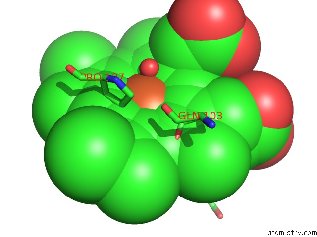
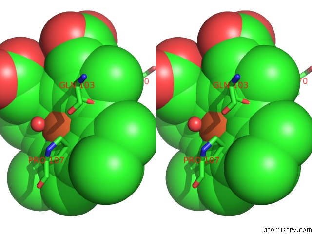
Iron binding site 1 out of 4 in 4k3i
Go back to
Iron binding site 1 out
of 4 in the Crystal Structure of the Quinol Form of Methylamine Dehydrogenase in Complex with the Diferrous Form of Maug, C2 Space Group
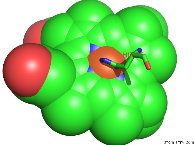
Mono view
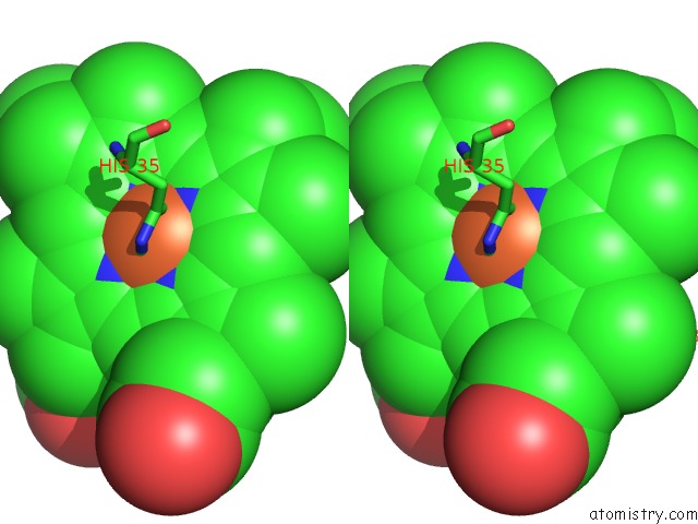
Stereo pair view
Mono view
Stereo pair view
A full contact list of Iron with other atoms in the Fe binding
site number 1 of Crystal Structure of the Quinol Form of Methylamine Dehydrogenase in Complex with the Diferrous Form of Maug, C2 Space Group within 5.0Å range:
|
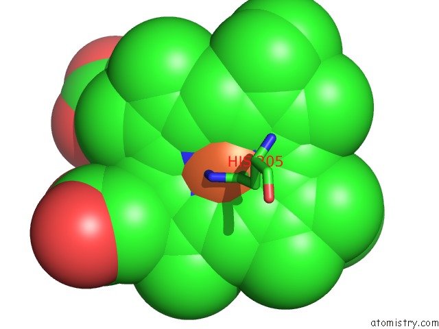
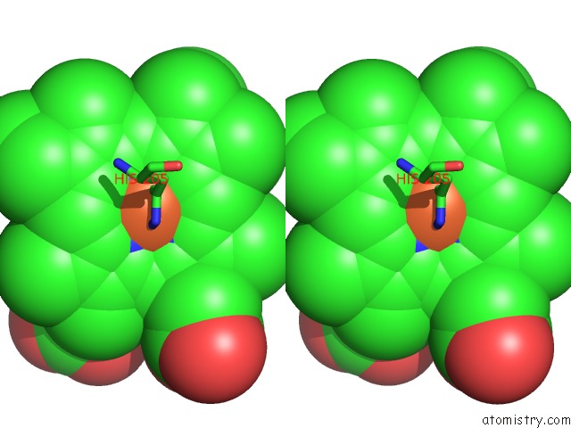
Iron binding site 2 out of 4 in 4k3i
Go back to
Iron binding site 2 out
of 4 in the Crystal Structure of the Quinol Form of Methylamine Dehydrogenase in Complex with the Diferrous Form of Maug, C2 Space Group
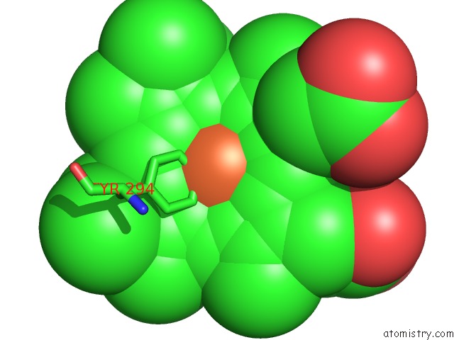
Mono view
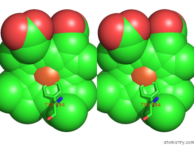
Stereo pair view
Mono view
Stereo pair view
A full contact list of Iron with other atoms in the Fe binding
site number 2 of Crystal Structure of the Quinol Form of Methylamine Dehydrogenase in Complex with the Diferrous Form of Maug, C2 Space Group within 5.0Å range:
|
Iron binding site 3 out of 4 in 4k3i
Go back to
Iron binding site 3 out
of 4 in the Crystal Structure of the Quinol Form of Methylamine Dehydrogenase in Complex with the Diferrous Form of Maug, C2 Space Group
Mono view
Stereo pair view
Mono view
Stereo pair view
A full contact list of Iron with other atoms in the Fe binding
site number 3 of Crystal Structure of the Quinol Form of Methylamine Dehydrogenase in Complex with the Diferrous Form of Maug, C2 Space Group within 5.0Å range:
|
Iron binding site 4 out of 4 in 4k3i
Go back to
Iron binding site 4 out
of 4 in the Crystal Structure of the Quinol Form of Methylamine Dehydrogenase in Complex with the Diferrous Form of Maug, C2 Space Group
Mono view
Stereo pair view
Mono view
Stereo pair view
A full contact list of Iron with other atoms in the Fe binding
site number 4 of Crystal Structure of the Quinol Form of Methylamine Dehydrogenase in Complex with the Diferrous Form of Maug, C2 Space Group within 5.0Å range:
|
Reference:
E.T.Yukl,
L.M.Jensen,
V.L.Davidson,
C.M.Wilmot.
Structures of Maug in Complex with Quinol and Quinone Madh. Acta Crystallogr.,Sect.F V. 69 738 2013.
ISSN: ESSN 1744-3091
PubMed: 23832199
DOI: 10.1107/S1744309113016539
Page generated: Mon Aug 5 05:00:28 2024
ISSN: ESSN 1744-3091
PubMed: 23832199
DOI: 10.1107/S1744309113016539
Last articles
Zn in 9MJ5Zn in 9HNW
Zn in 9G0L
Zn in 9FNE
Zn in 9DZN
Zn in 9E0I
Zn in 9D32
Zn in 9DAK
Zn in 8ZXC
Zn in 8ZUF