Iron »
PDB 4k5e-4kf2 »
4k8f »
Iron in PDB 4k8f: Structure of the Heme Domain of Cooa From Rhodospirillum Rubrum
Protein crystallography data
The structure of Structure of the Heme Domain of Cooa From Rhodospirillum Rubrum, PDB code: 4k8f
was solved by
M.Kuchinskas,
H.Li,
T.L.Poulos,
with X-Ray Crystallography technique. A brief refinement statistics is given in the table below:
| Resolution Low / High (Å) | 35.62 / 2.70 |
| Space group | P 41 |
| Cell size a, b, c (Å), α, β, γ (°) | 71.230, 71.230, 143.733, 90.00, 90.00, 90.00 |
| R / Rfree (%) | 19.7 / 26.3 |
Iron Binding Sites:
The binding sites of Iron atom in the Structure of the Heme Domain of Cooa From Rhodospirillum Rubrum
(pdb code 4k8f). This binding sites where shown within
5.0 Angstroms radius around Iron atom.
In total 4 binding sites of Iron where determined in the Structure of the Heme Domain of Cooa From Rhodospirillum Rubrum, PDB code: 4k8f:
Jump to Iron binding site number: 1; 2; 3; 4;
In total 4 binding sites of Iron where determined in the Structure of the Heme Domain of Cooa From Rhodospirillum Rubrum, PDB code: 4k8f:
Jump to Iron binding site number: 1; 2; 3; 4;
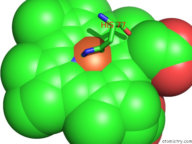
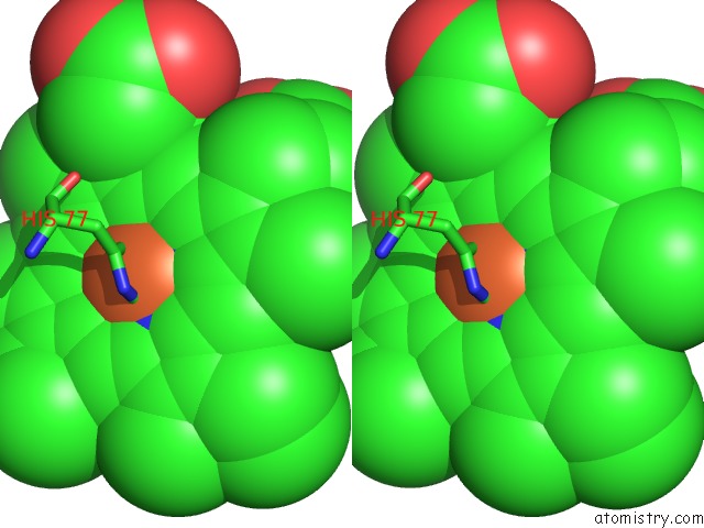
Iron binding site 1 out of 4 in 4k8f
Go back to
Iron binding site 1 out
of 4 in the Structure of the Heme Domain of Cooa From Rhodospirillum Rubrum
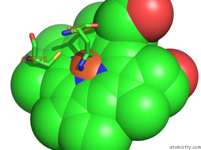
Mono view
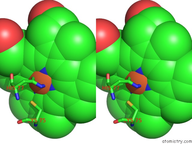
Stereo pair view
Mono view
Stereo pair view
A full contact list of Iron with other atoms in the Fe binding
site number 1 of Structure of the Heme Domain of Cooa From Rhodospirillum Rubrum within 5.0Å range:
|
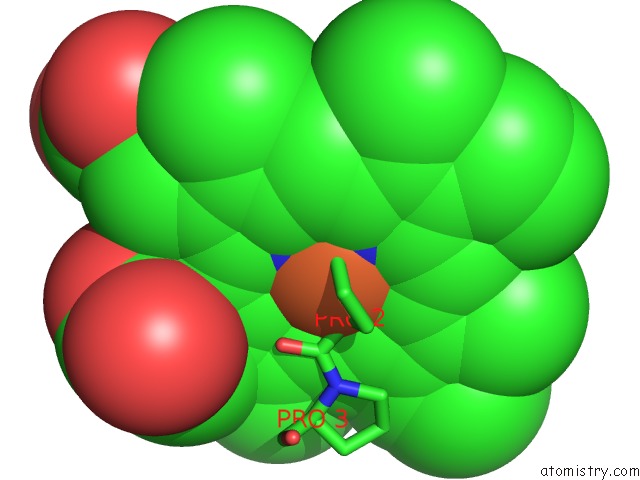
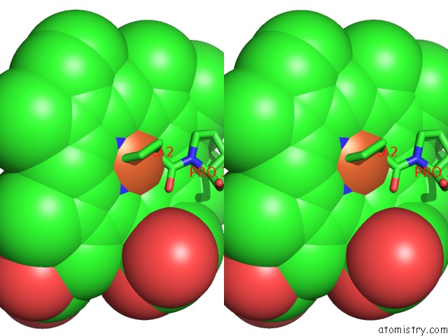
Iron binding site 2 out of 4 in 4k8f
Go back to
Iron binding site 2 out
of 4 in the Structure of the Heme Domain of Cooa From Rhodospirillum Rubrum
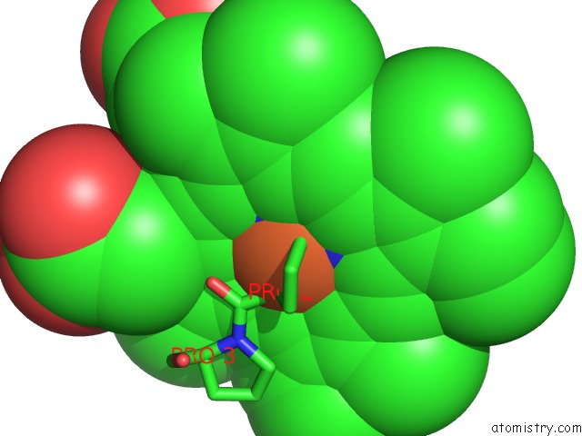
Mono view
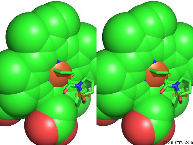
Stereo pair view
Mono view
Stereo pair view
A full contact list of Iron with other atoms in the Fe binding
site number 2 of Structure of the Heme Domain of Cooa From Rhodospirillum Rubrum within 5.0Å range:
|
Iron binding site 3 out of 4 in 4k8f
Go back to
Iron binding site 3 out
of 4 in the Structure of the Heme Domain of Cooa From Rhodospirillum Rubrum
Mono view
Stereo pair view
Mono view
Stereo pair view
A full contact list of Iron with other atoms in the Fe binding
site number 3 of Structure of the Heme Domain of Cooa From Rhodospirillum Rubrum within 5.0Å range:
|
Iron binding site 4 out of 4 in 4k8f
Go back to
Iron binding site 4 out
of 4 in the Structure of the Heme Domain of Cooa From Rhodospirillum Rubrum
Mono view
Stereo pair view
Mono view
Stereo pair view
A full contact list of Iron with other atoms in the Fe binding
site number 4 of Structure of the Heme Domain of Cooa From Rhodospirillum Rubrum within 5.0Å range:
|
Reference:
M.Kuchinskas,
H.Li,
M.Conrad,
G.Roberts,
T.L.Poulos.
The Role of the Dna-Binding Domains in Cooa Activation. Biochemistry V. 45 7148 2006.
ISSN: ISSN 0006-2960
PubMed: 16752905
DOI: 10.1021/BI052609O
Page generated: Mon Aug 5 05:17:31 2024
ISSN: ISSN 0006-2960
PubMed: 16752905
DOI: 10.1021/BI052609O
Last articles
F in 7REKF in 7REL
F in 7RE2
F in 7REE
F in 7RE1
F in 7RE0
F in 7RDY
F in 7RDX
F in 7RD7
F in 7RD6