Iron »
PDB 4kib-4kx6 »
4kmg »
Iron in PDB 4kmg: Crystal Structure of Cytochrome C6B From Synechococcus Sp. WH8102
Protein crystallography data
The structure of Crystal Structure of Cytochrome C6B From Synechococcus Sp. WH8102, PDB code: 4kmg
was solved by
P.Zatwarnicki,
S.Krzywda,
J.Barciszewski,
M.Jaskolski,
A.Szczepaniak,
with X-Ray Crystallography technique. A brief refinement statistics is given in the table below:
| Resolution Low / High (Å) | 27.95 / 1.40 |
| Space group | C 1 2 1 |
| Cell size a, b, c (Å), α, β, γ (°) | 106.110, 28.980, 24.680, 90.00, 92.30, 90.00 |
| R / Rfree (%) | 12 / 18.1 |
Other elements in 4kmg:
The structure of Crystal Structure of Cytochrome C6B From Synechococcus Sp. WH8102 also contains other interesting chemical elements:
| Sodium | (Na) | 1 atom |
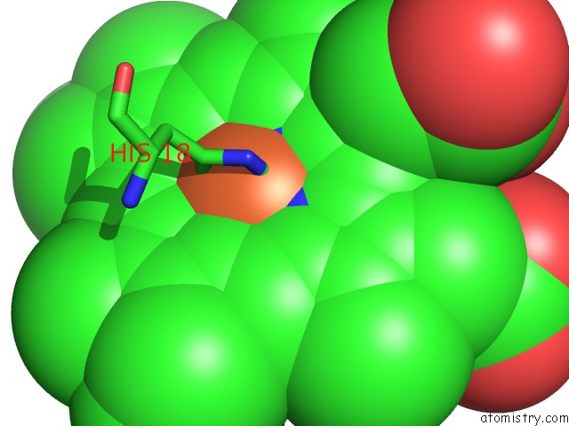
Iron Binding Sites:
The binding sites of Iron atom in the Crystal Structure of Cytochrome C6B From Synechococcus Sp. WH8102
(pdb code 4kmg). This binding sites where shown within
5.0 Angstroms radius around Iron atom.
In total only one binding site of Iron was determined in the Crystal Structure of Cytochrome C6B From Synechococcus Sp. WH8102, PDB code: 4kmg:
In total only one binding site of Iron was determined in the Crystal Structure of Cytochrome C6B From Synechococcus Sp. WH8102, PDB code: 4kmg:
Iron binding site 1 out of 1 in 4kmg
Go back to
Iron binding site 1 out
of 1 in the Crystal Structure of Cytochrome C6B From Synechococcus Sp. WH8102
Mono view

Stereo pair view
Mono view
Stereo pair view
A full contact list of Iron with other atoms in the Fe binding
site number 1 of Crystal Structure of Cytochrome C6B From Synechococcus Sp. WH8102 within 5.0Å range:
|
Reference:
P.Zatwarnicki,
J.Barciszewski,
S.Krzywda,
M.Jaskolski,
P.Kolesinski,
A.Szczepaniak.
Cytochrome C6B of Synechococcus Sp. Wh 8102 - Crystal Structure and Basic Properties of Novel C6-Like Family Representative. Biochem.Biophys.Res.Commun. V. 443 1131 2014.
ISSN: ISSN 0006-291X
PubMed: 24216109
DOI: 10.1016/J.BBRC.2013.10.167
Page generated: Tue Aug 5 12:00:27 2025
ISSN: ISSN 0006-291X
PubMed: 24216109
DOI: 10.1016/J.BBRC.2013.10.167
Last articles
Fe in 4S01Fe in 4S00
Fe in 4RZY
Fe in 4RZZ
Fe in 4RYZ
Fe in 4RYY
Fe in 4RYX
Fe in 4RKM
Fe in 4RWM
Fe in 4RXN