Iron »
PDB 4kib-4kx6 »
4kpa »
Iron in PDB 4kpa: Crystal Structure of Cytochrome P450 Bm-3 in Complex with N- Palmitoylglycine (Npg)
Enzymatic activity of Crystal Structure of Cytochrome P450 Bm-3 in Complex with N- Palmitoylglycine (Npg)
All present enzymatic activity of Crystal Structure of Cytochrome P450 Bm-3 in Complex with N- Palmitoylglycine (Npg):
1.14.14.1;
1.14.14.1;
Protein crystallography data
The structure of Crystal Structure of Cytochrome P450 Bm-3 in Complex with N- Palmitoylglycine (Npg), PDB code: 4kpa
was solved by
G.A.Amadeo,
J.Catalano,
A.E.Mcdermott,
L.Tong,
with X-Ray Crystallography technique. A brief refinement statistics is given in the table below:
| Resolution Low / High (Å) | 29.66 / 2.00 |
| Space group | P 21 21 2 |
| Cell size a, b, c (Å), α, β, γ (°) | 188.700, 59.320, 56.240, 90.00, 90.00, 90.00 |
| R / Rfree (%) | 19.4 / 23 |
Iron Binding Sites:
The binding sites of Iron atom in the Crystal Structure of Cytochrome P450 Bm-3 in Complex with N- Palmitoylglycine (Npg)
(pdb code 4kpa). This binding sites where shown within
5.0 Angstroms radius around Iron atom.
In total only one binding site of Iron was determined in the Crystal Structure of Cytochrome P450 Bm-3 in Complex with N- Palmitoylglycine (Npg), PDB code: 4kpa:
In total only one binding site of Iron was determined in the Crystal Structure of Cytochrome P450 Bm-3 in Complex with N- Palmitoylglycine (Npg), PDB code: 4kpa:
Iron binding site 1 out of 1 in 4kpa
Go back to
Iron binding site 1 out
of 1 in the Crystal Structure of Cytochrome P450 Bm-3 in Complex with N- Palmitoylglycine (Npg)
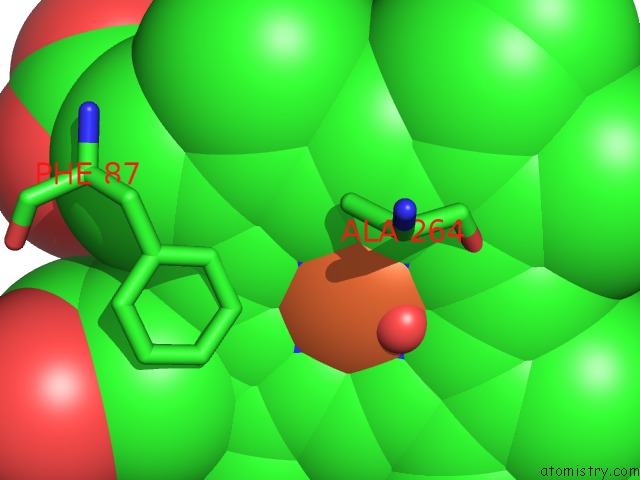
Mono view
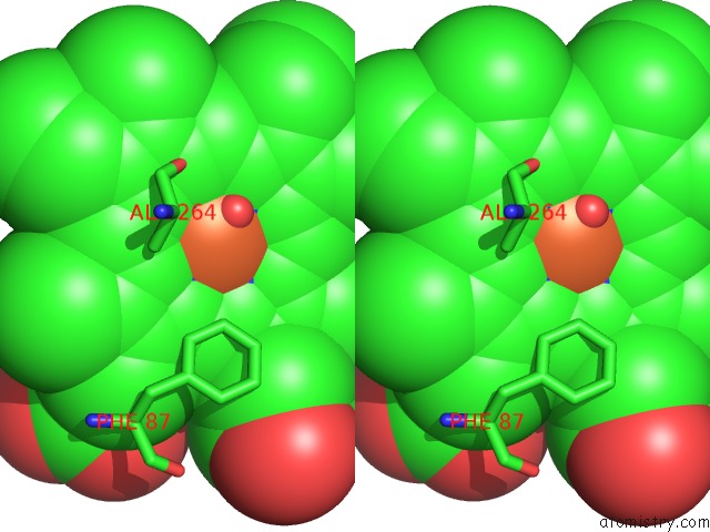
Stereo pair view
Mono view
Stereo pair view
A full contact list of Iron with other atoms in the Fe binding
site number 1 of Crystal Structure of Cytochrome P450 Bm-3 in Complex with N- Palmitoylglycine (Npg) within 5.0Å range:
|
Reference:
J.Catalano,
K.Sadre-Bazzaz,
G.A.Amodeo,
L.Tong,
A.Mcdermott.
Structural Evidence: A Single Charged Residue Affects Substrate Binding in Cytochrome P450 Bm-3. Biochemistry V. 52 6807 2013.
ISSN: ISSN 0006-2960
PubMed: 23829560
DOI: 10.1021/BI4000645
Page generated: Tue Aug 5 12:11:32 2025
ISSN: ISSN 0006-2960
PubMed: 23829560
DOI: 10.1021/BI4000645
Last articles
Fe in 4RYXFe in 4RKM
Fe in 4RWM
Fe in 4RXN
Fe in 4RUI
Fe in 4RVY
Fe in 4RKN
Fe in 4RSZ
Fe in 4RU3
Fe in 4RTB