Iron »
PDB 4kib-4kx6 »
4ktj »
Iron in PDB 4ktj: Crystal Structure of Mycobacterium Tuberculosis CYP121 in Complex with 4-(3-Amino-1H-Pyrazol-4-Yl)Phenol
Protein crystallography data
The structure of Crystal Structure of Mycobacterium Tuberculosis CYP121 in Complex with 4-(3-Amino-1H-Pyrazol-4-Yl)Phenol, PDB code: 4ktj
was solved by
S.A.Hudson,
with X-Ray Crystallography technique. A brief refinement statistics is given in the table below:
| Resolution Low / High (Å) | 33.62 / 1.35 |
| Space group | P 65 2 2 |
| Cell size a, b, c (Å), α, β, γ (°) | 78.030, 78.030, 265.161, 90.00, 90.00, 120.00 |
| R / Rfree (%) | 16.5 / 19.5 |
Iron Binding Sites:
The binding sites of Iron atom in the Crystal Structure of Mycobacterium Tuberculosis CYP121 in Complex with 4-(3-Amino-1H-Pyrazol-4-Yl)Phenol
(pdb code 4ktj). This binding sites where shown within
5.0 Angstroms radius around Iron atom.
In total only one binding site of Iron was determined in the Crystal Structure of Mycobacterium Tuberculosis CYP121 in Complex with 4-(3-Amino-1H-Pyrazol-4-Yl)Phenol, PDB code: 4ktj:
In total only one binding site of Iron was determined in the Crystal Structure of Mycobacterium Tuberculosis CYP121 in Complex with 4-(3-Amino-1H-Pyrazol-4-Yl)Phenol, PDB code: 4ktj:
Iron binding site 1 out of 1 in 4ktj
Go back to
Iron binding site 1 out
of 1 in the Crystal Structure of Mycobacterium Tuberculosis CYP121 in Complex with 4-(3-Amino-1H-Pyrazol-4-Yl)Phenol
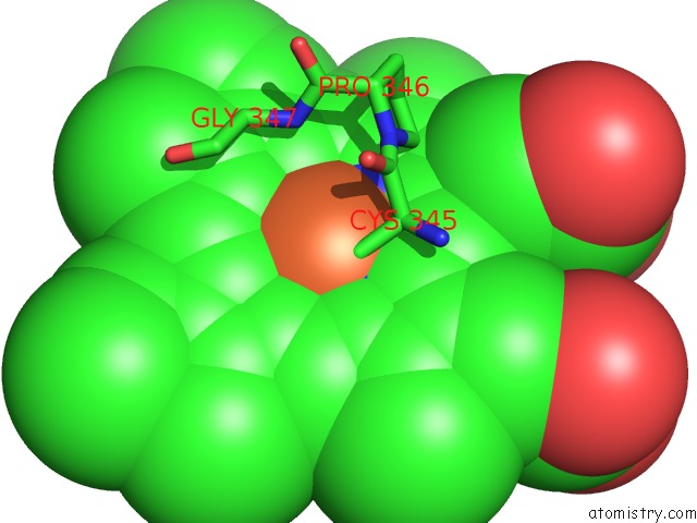
Mono view
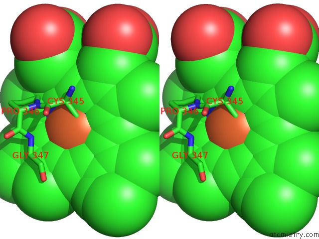
Stereo pair view
Mono view
Stereo pair view
A full contact list of Iron with other atoms in the Fe binding
site number 1 of Crystal Structure of Mycobacterium Tuberculosis CYP121 in Complex with 4-(3-Amino-1H-Pyrazol-4-Yl)Phenol within 5.0Å range:
|
Reference:
S.A.Hudson,
S.Surade,
A.G.Coyne,
K.J.Mclean,
D.Leys,
A.W.Munro,
C.Abell.
Overcoming the Limitations of Fragment Merging: Rescuing A Strained Merged Fragment Series Targeting Mycobacterium Tuberculosis CYP121. Chemmedchem V. 8 1451 2013.
ISSN: ISSN 1860-7179
PubMed: 23788280
DOI: 10.1002/CMDC.201300219
Page generated: Mon Aug 5 05:35:59 2024
ISSN: ISSN 1860-7179
PubMed: 23788280
DOI: 10.1002/CMDC.201300219
Last articles
Zn in 9MJ5Zn in 9HNW
Zn in 9G0L
Zn in 9FNE
Zn in 9DZN
Zn in 9E0I
Zn in 9D32
Zn in 9DAK
Zn in 8ZXC
Zn in 8ZUF