Iron »
PDB 4l0d-4lji »
4l2m »
Iron in PDB 4l2m: Crystal Structure of the 2/2 Hemoglobin From Synechococcus Sp. Pcc 7002 in the Cyanomet State and with Covalently Attached Heme
Protein crystallography data
The structure of Crystal Structure of the 2/2 Hemoglobin From Synechococcus Sp. Pcc 7002 in the Cyanomet State and with Covalently Attached Heme, PDB code: 4l2m
was solved by
B.B.Wenke,
J.L.Schlessman,
A.Heroux,
J.T.J.Lecomte,
with X-Ray Crystallography technique. A brief refinement statistics is given in the table below:
| Resolution Low / High (Å) | 34.19 / 2.25 |
| Space group | P 1 21 1 |
| Cell size a, b, c (Å), α, β, γ (°) | 50.622, 40.951, 66.461, 90.00, 111.21, 90.00 |
| R / Rfree (%) | 21.8 / 28.8 |
Iron Binding Sites:
The binding sites of Iron atom in the Crystal Structure of the 2/2 Hemoglobin From Synechococcus Sp. Pcc 7002 in the Cyanomet State and with Covalently Attached Heme
(pdb code 4l2m). This binding sites where shown within
5.0 Angstroms radius around Iron atom.
In total 2 binding sites of Iron where determined in the Crystal Structure of the 2/2 Hemoglobin From Synechococcus Sp. Pcc 7002 in the Cyanomet State and with Covalently Attached Heme, PDB code: 4l2m:
Jump to Iron binding site number: 1; 2;
In total 2 binding sites of Iron where determined in the Crystal Structure of the 2/2 Hemoglobin From Synechococcus Sp. Pcc 7002 in the Cyanomet State and with Covalently Attached Heme, PDB code: 4l2m:
Jump to Iron binding site number: 1; 2;
Iron binding site 1 out of 2 in 4l2m
Go back to
Iron binding site 1 out
of 2 in the Crystal Structure of the 2/2 Hemoglobin From Synechococcus Sp. Pcc 7002 in the Cyanomet State and with Covalently Attached Heme
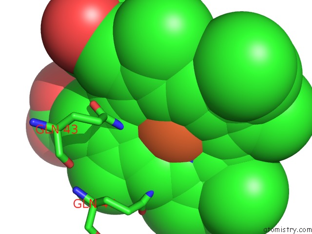
Mono view
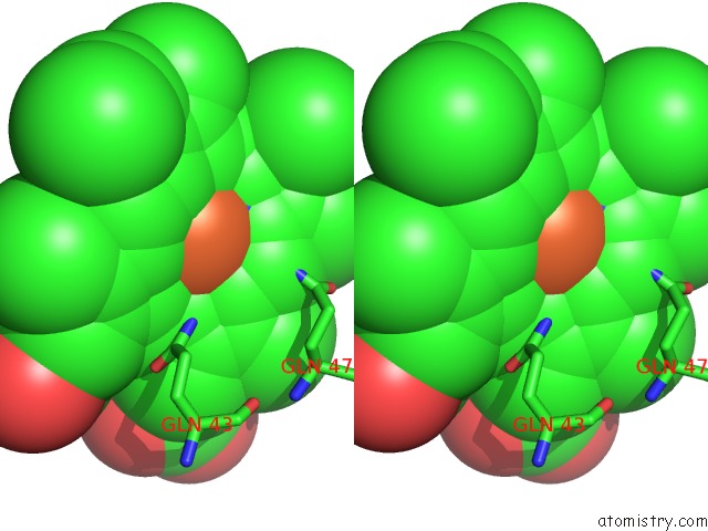
Stereo pair view
Mono view
Stereo pair view
A full contact list of Iron with other atoms in the Fe binding
site number 1 of Crystal Structure of the 2/2 Hemoglobin From Synechococcus Sp. Pcc 7002 in the Cyanomet State and with Covalently Attached Heme within 5.0Å range:
|
Iron binding site 2 out of 2 in 4l2m
Go back to
Iron binding site 2 out
of 2 in the Crystal Structure of the 2/2 Hemoglobin From Synechococcus Sp. Pcc 7002 in the Cyanomet State and with Covalently Attached Heme
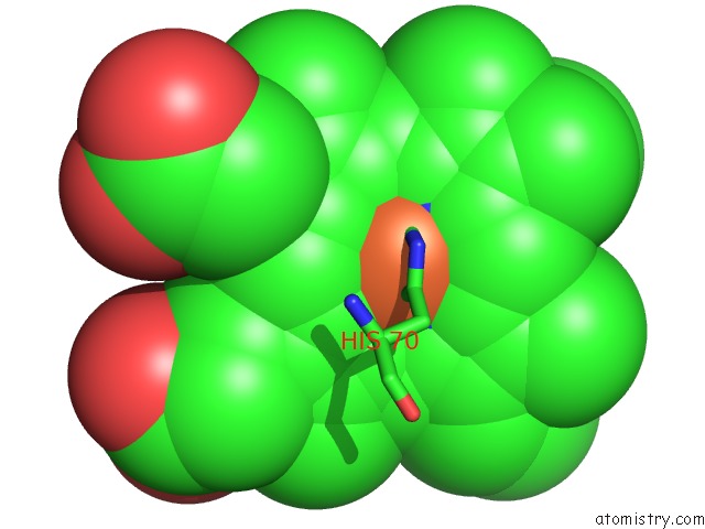
Mono view
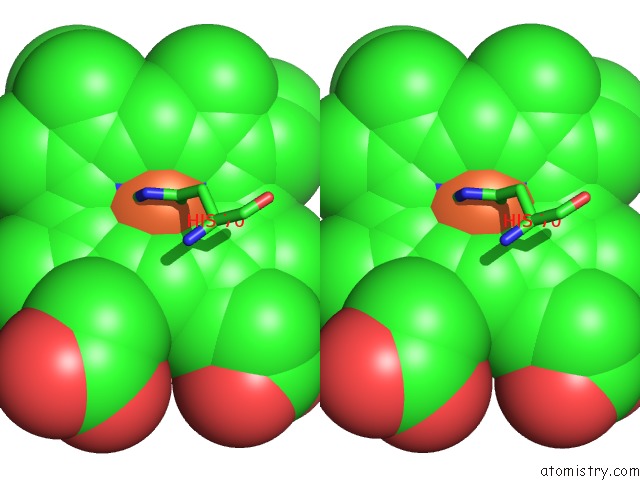
Stereo pair view
Mono view
Stereo pair view
A full contact list of Iron with other atoms in the Fe binding
site number 2 of Crystal Structure of the 2/2 Hemoglobin From Synechococcus Sp. Pcc 7002 in the Cyanomet State and with Covalently Attached Heme within 5.0Å range:
|
Reference:
B.B.Wenke,
J.T.Lecomte,
A.Heroux,
J.L.Schlessman.
The 2/2 Hemoglobin From the Cyanobacterium Synechococcus Sp. Pcc 7002 with Covalently Attached Heme: Comparison of X-Ray and uc(Nmr) Structures. Proteins V. 82 528 2014.
ISSN: ISSN 0887-3585
PubMed: 23999883
DOI: 10.1002/PROT.24409
Page generated: Tue Aug 5 12:22:03 2025
ISSN: ISSN 0887-3585
PubMed: 23999883
DOI: 10.1002/PROT.24409
Last articles
Fe in 4S3BFe in 4S3A
Fe in 4S39
Fe in 4S38
Fe in 4S2A
Fe in 4S26
Fe in 4S36
Fe in 4S28
Fe in 4S23
Fe in 4S29