Iron »
PDB 4l0d-4lji »
4l3h »
Iron in PDB 4l3h: Crystal Structure of the E113Q-Maug/Pre-Methylamine Dehydrogenase Complex After Treatment with Hydrogen Peroxide
Enzymatic activity of Crystal Structure of the E113Q-Maug/Pre-Methylamine Dehydrogenase Complex After Treatment with Hydrogen Peroxide
All present enzymatic activity of Crystal Structure of the E113Q-Maug/Pre-Methylamine Dehydrogenase Complex After Treatment with Hydrogen Peroxide:
1.4.99.3;
1.4.99.3;
Protein crystallography data
The structure of Crystal Structure of the E113Q-Maug/Pre-Methylamine Dehydrogenase Complex After Treatment with Hydrogen Peroxide, PDB code: 4l3h
was solved by
E.T.Yukl,
C.M.Wilmot,
with X-Ray Crystallography technique. A brief refinement statistics is given in the table below:
| Resolution Low / High (Å) | 44.49 / 1.79 |
| Space group | P 1 |
| Cell size a, b, c (Å), α, β, γ (°) | 55.530, 83.520, 107.780, 109.94, 91.54, 105.78 |
| R / Rfree (%) | 15.4 / 19.8 |
Other elements in 4l3h:
The structure of Crystal Structure of the E113Q-Maug/Pre-Methylamine Dehydrogenase Complex After Treatment with Hydrogen Peroxide also contains other interesting chemical elements:
| Calcium | (Ca) | 2 atoms |
| Sodium | (Na) | 4 atoms |
Iron Binding Sites:
The binding sites of Iron atom in the Crystal Structure of the E113Q-Maug/Pre-Methylamine Dehydrogenase Complex After Treatment with Hydrogen Peroxide
(pdb code 4l3h). This binding sites where shown within
5.0 Angstroms radius around Iron atom.
In total 4 binding sites of Iron where determined in the Crystal Structure of the E113Q-Maug/Pre-Methylamine Dehydrogenase Complex After Treatment with Hydrogen Peroxide, PDB code: 4l3h:
Jump to Iron binding site number: 1; 2; 3; 4;
In total 4 binding sites of Iron where determined in the Crystal Structure of the E113Q-Maug/Pre-Methylamine Dehydrogenase Complex After Treatment with Hydrogen Peroxide, PDB code: 4l3h:
Jump to Iron binding site number: 1; 2; 3; 4;
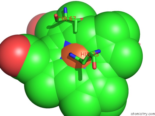
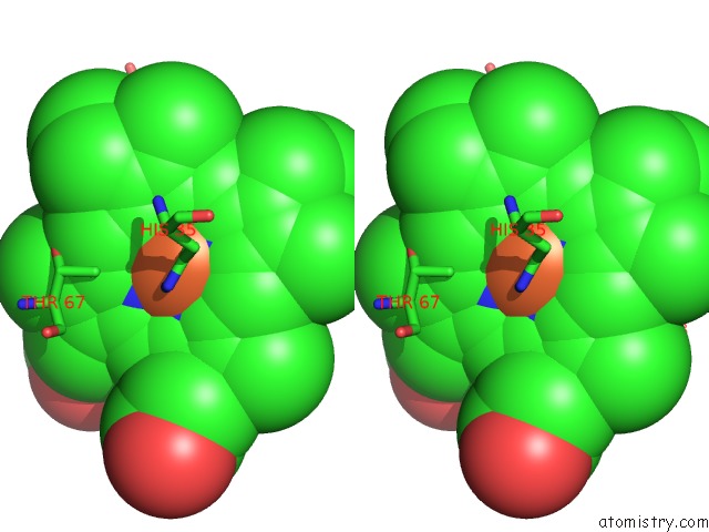
Iron binding site 1 out of 4 in 4l3h
Go back to
Iron binding site 1 out
of 4 in the Crystal Structure of the E113Q-Maug/Pre-Methylamine Dehydrogenase Complex After Treatment with Hydrogen Peroxide
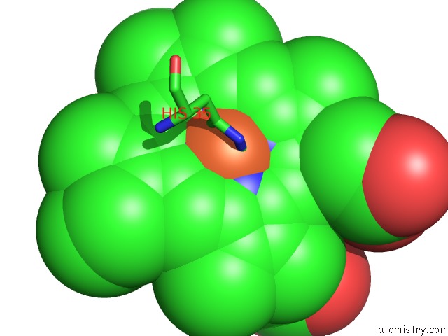
Mono view
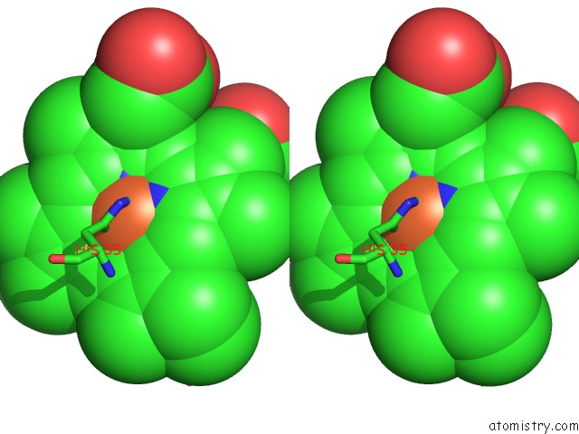
Stereo pair view
Mono view
Stereo pair view
A full contact list of Iron with other atoms in the Fe binding
site number 1 of Crystal Structure of the E113Q-Maug/Pre-Methylamine Dehydrogenase Complex After Treatment with Hydrogen Peroxide within 5.0Å range:
|
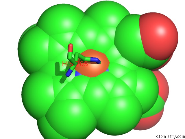
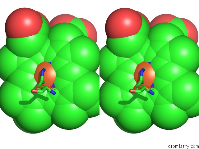
Iron binding site 2 out of 4 in 4l3h
Go back to
Iron binding site 2 out
of 4 in the Crystal Structure of the E113Q-Maug/Pre-Methylamine Dehydrogenase Complex After Treatment with Hydrogen Peroxide
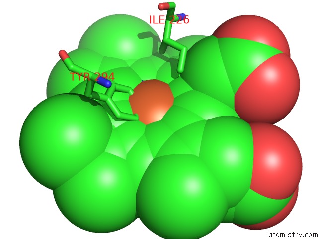
Mono view
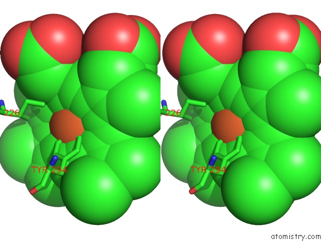
Stereo pair view
Mono view
Stereo pair view
A full contact list of Iron with other atoms in the Fe binding
site number 2 of Crystal Structure of the E113Q-Maug/Pre-Methylamine Dehydrogenase Complex After Treatment with Hydrogen Peroxide within 5.0Å range:
|
Iron binding site 3 out of 4 in 4l3h
Go back to
Iron binding site 3 out
of 4 in the Crystal Structure of the E113Q-Maug/Pre-Methylamine Dehydrogenase Complex After Treatment with Hydrogen Peroxide
Mono view
Stereo pair view
Mono view
Stereo pair view
A full contact list of Iron with other atoms in the Fe binding
site number 3 of Crystal Structure of the E113Q-Maug/Pre-Methylamine Dehydrogenase Complex After Treatment with Hydrogen Peroxide within 5.0Å range:
|
Iron binding site 4 out of 4 in 4l3h
Go back to
Iron binding site 4 out
of 4 in the Crystal Structure of the E113Q-Maug/Pre-Methylamine Dehydrogenase Complex After Treatment with Hydrogen Peroxide
Mono view
Stereo pair view
Mono view
Stereo pair view
A full contact list of Iron with other atoms in the Fe binding
site number 4 of Crystal Structure of the E113Q-Maug/Pre-Methylamine Dehydrogenase Complex After Treatment with Hydrogen Peroxide within 5.0Å range:
|
Reference:
N.Abu Tarboush,
E.T.Yukl,
S.Shin,
M.Feng,
C.M.Wilmot,
V.L.Davidson.
Carboxyl Group of GLU113 Is Required For Stabilization of the Diferrous and Bis-Fe(IV) States of Maug. Biochemistry V. 52 6358 2013.
ISSN: ISSN 0006-2960
PubMed: 23952537
DOI: 10.1021/BI400905S
Page generated: Mon Aug 5 06:05:29 2024
ISSN: ISSN 0006-2960
PubMed: 23952537
DOI: 10.1021/BI400905S
Last articles
Zn in 9MJ5Zn in 9HNW
Zn in 9G0L
Zn in 9FNE
Zn in 9DZN
Zn in 9E0I
Zn in 9D32
Zn in 9DAK
Zn in 8ZXC
Zn in 8ZUF