Iron »
PDB 4n4j-4nji »
4n59 »
Iron in PDB 4n59: The Crystal Structure of Pectocin M2 at 2.3 Angstroms
Protein crystallography data
The structure of The Crystal Structure of Pectocin M2 at 2.3 Angstroms, PDB code: 4n59
was solved by
K.Zeth,
R.Grinter,
A.W.Roszak,
R.J.Cogdell,
D.Walker,
with X-Ray Crystallography technique. A brief refinement statistics is given in the table below:
| Resolution Low / High (Å) | 44.52 / 2.30 |
| Space group | P 1 21 1 |
| Cell size a, b, c (Å), α, β, γ (°) | 44.649, 116.745, 60.790, 90.00, 94.96, 90.00 |
| R / Rfree (%) | 20.6 / 27.4 |
Other elements in 4n59:
The structure of The Crystal Structure of Pectocin M2 at 2.3 Angstroms also contains other interesting chemical elements:
| Chlorine | (Cl) | 1 atom |
Iron Binding Sites:
The binding sites of Iron atom in the The Crystal Structure of Pectocin M2 at 2.3 Angstroms
(pdb code 4n59). This binding sites where shown within
5.0 Angstroms radius around Iron atom.
In total 4 binding sites of Iron where determined in the The Crystal Structure of Pectocin M2 at 2.3 Angstroms, PDB code: 4n59:
Jump to Iron binding site number: 1; 2; 3; 4;
In total 4 binding sites of Iron where determined in the The Crystal Structure of Pectocin M2 at 2.3 Angstroms, PDB code: 4n59:
Jump to Iron binding site number: 1; 2; 3; 4;
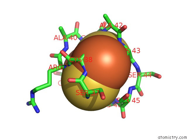
Iron binding site 1 out of 4 in 4n59
Go back to
Iron binding site 1 out
of 4 in the The Crystal Structure of Pectocin M2 at 2.3 Angstroms
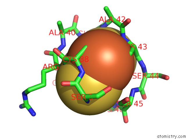
Mono view
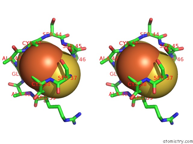
Stereo pair view
Mono view
Stereo pair view
A full contact list of Iron with other atoms in the Fe binding
site number 1 of The Crystal Structure of Pectocin M2 at 2.3 Angstroms within 5.0Å range:
|
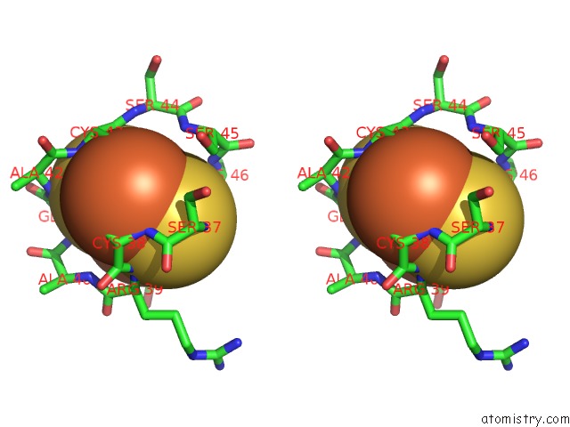
Iron binding site 2 out of 4 in 4n59
Go back to
Iron binding site 2 out
of 4 in the The Crystal Structure of Pectocin M2 at 2.3 Angstroms
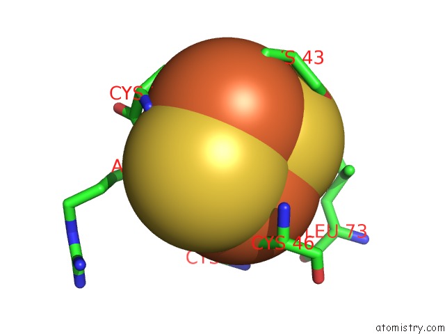
Mono view
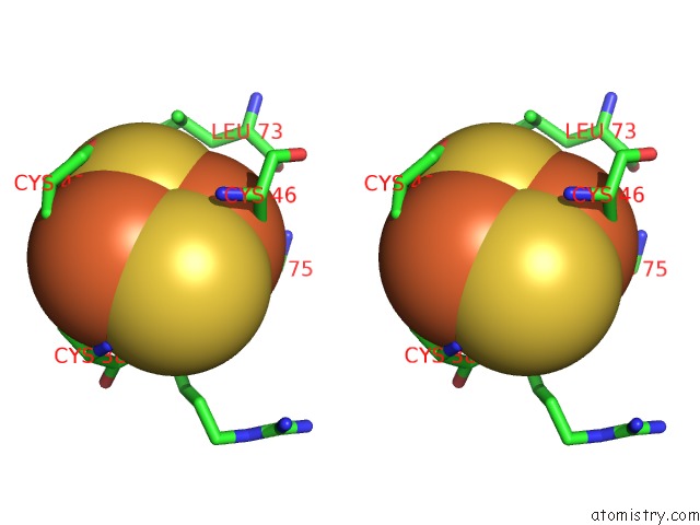
Stereo pair view
Mono view
Stereo pair view
A full contact list of Iron with other atoms in the Fe binding
site number 2 of The Crystal Structure of Pectocin M2 at 2.3 Angstroms within 5.0Å range:
|
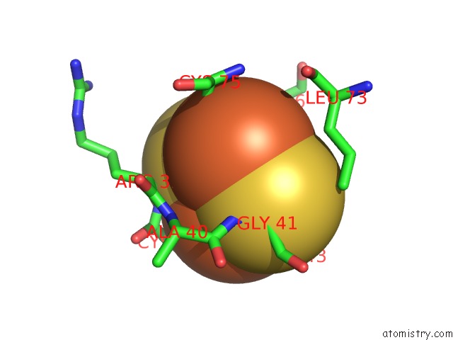
Iron binding site 3 out of 4 in 4n59
Go back to
Iron binding site 3 out
of 4 in the The Crystal Structure of Pectocin M2 at 2.3 Angstroms
Mono view
Stereo pair view
Mono view
Stereo pair view
A full contact list of Iron with other atoms in the Fe binding
site number 3 of The Crystal Structure of Pectocin M2 at 2.3 Angstroms within 5.0Å range:
|
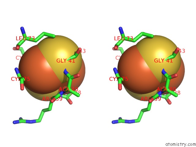
Iron binding site 4 out of 4 in 4n59
Go back to
Iron binding site 4 out
of 4 in the The Crystal Structure of Pectocin M2 at 2.3 Angstroms
Mono view
Stereo pair view
Mono view
Stereo pair view
A full contact list of Iron with other atoms in the Fe binding
site number 4 of The Crystal Structure of Pectocin M2 at 2.3 Angstroms within 5.0Å range:
|
Reference:
R.Grinter,
I.Josts,
K.Zeth,
A.W.Roszak,
L.C.Mccaughey,
R.J.Cogdell,
J.J.Milner,
S.M.Kelly,
O.Byron,
D.Walker.
Structure of the Atypical Bacteriocin Pectocin M2 Implies A Novel Mechanism of Protein Uptake. Mol.Microbiol. V. 93 234 2014.
ISSN: ISSN 0950-382X
PubMed: 24865810
DOI: 10.1111/MMI.12655
Page generated: Mon Aug 5 07:09:23 2024
ISSN: ISSN 0950-382X
PubMed: 24865810
DOI: 10.1111/MMI.12655
Last articles
Zn in 9J0NZn in 9J0O
Zn in 9J0P
Zn in 9FJX
Zn in 9EKB
Zn in 9C0F
Zn in 9CAH
Zn in 9CH0
Zn in 9CH3
Zn in 9CH1