Iron »
PDB 4n4j-4nji »
4nao »
Iron in PDB 4nao: Crystal Structure of Eash
Protein crystallography data
The structure of Crystal Structure of Eash, PDB code: 4nao
was solved by
R.Janke,
J.Havemann,
D.Vogel,
U.Keller,
B.Loll,
with X-Ray Crystallography technique. A brief refinement statistics is given in the table below:
| Resolution Low / High (Å) | 36.33 / 1.65 |
| Space group | P 43 21 2 |
| Cell size a, b, c (Å), α, β, γ (°) | 91.304, 91.304, 79.189, 90.00, 90.00, 90.00 |
| R / Rfree (%) | 15.7 / 18.2 |
Other elements in 4nao:
The structure of Crystal Structure of Eash also contains other interesting chemical elements:
| Sodium | (Na) | 1 atom |
Iron Binding Sites:
The binding sites of Iron atom in the Crystal Structure of Eash
(pdb code 4nao). This binding sites where shown within
5.0 Angstroms radius around Iron atom.
In total only one binding site of Iron was determined in the Crystal Structure of Eash, PDB code: 4nao:
In total only one binding site of Iron was determined in the Crystal Structure of Eash, PDB code: 4nao:
Iron binding site 1 out of 1 in 4nao
Go back to
Iron binding site 1 out
of 1 in the Crystal Structure of Eash
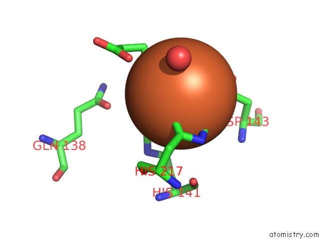
Mono view
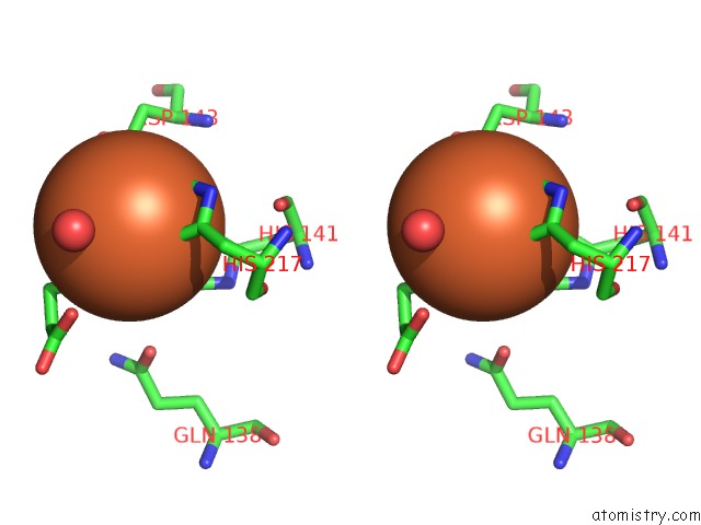
Stereo pair view
Mono view
Stereo pair view
A full contact list of Iron with other atoms in the Fe binding
site number 1 of Crystal Structure of Eash within 5.0Å range:
|
Reference:
J.Havemann,
D.Vogel,
B.Loll,
U.Keller.
Cyclolization of D-Lysergic Acid Alkaloid Peptides. Chem.Biol. V. 21 146 2014.
ISSN: ISSN 1074-5521
PubMed: 24361048
DOI: 10.1016/J.CHEMBIOL.2013.11.008
Page generated: Tue Aug 5 13:03:36 2025
ISSN: ISSN 1074-5521
PubMed: 24361048
DOI: 10.1016/J.CHEMBIOL.2013.11.008
Last articles
Fe in 4TO9Fe in 4TOB
Fe in 4TKV
Fe in 4TKU
Fe in 4TNK
Fe in 4TNJ
Fe in 4TNI
Fe in 4TNH
Fe in 4TLF
Fe in 4TN7