Iron »
PDB 4o1w-4oxf »
4o5v »
Iron in PDB 4o5v: Crystal Structure of T. Acidophilum Ider
Protein crystallography data
The structure of Crystal Structure of T. Acidophilum Ider, PDB code: 4o5v
was solved by
J.Y.Lee,
H.K.Yeo,
with X-Ray Crystallography technique. A brief refinement statistics is given in the table below:
| Resolution Low / High (Å) | 34.17 / 2.10 |
| Space group | P 21 21 2 |
| Cell size a, b, c (Å), α, β, γ (°) | 61.198, 84.978, 47.078, 90.00, 90.00, 90.00 |
| R / Rfree (%) | 18.2 / 22.5 |
Iron Binding Sites:
The binding sites of Iron atom in the Crystal Structure of T. Acidophilum Ider
(pdb code 4o5v). This binding sites where shown within
5.0 Angstroms radius around Iron atom.
In total 2 binding sites of Iron where determined in the Crystal Structure of T. Acidophilum Ider, PDB code: 4o5v:
Jump to Iron binding site number: 1; 2;
In total 2 binding sites of Iron where determined in the Crystal Structure of T. Acidophilum Ider, PDB code: 4o5v:
Jump to Iron binding site number: 1; 2;
Iron binding site 1 out of 2 in 4o5v
Go back to
Iron binding site 1 out
of 2 in the Crystal Structure of T. Acidophilum Ider
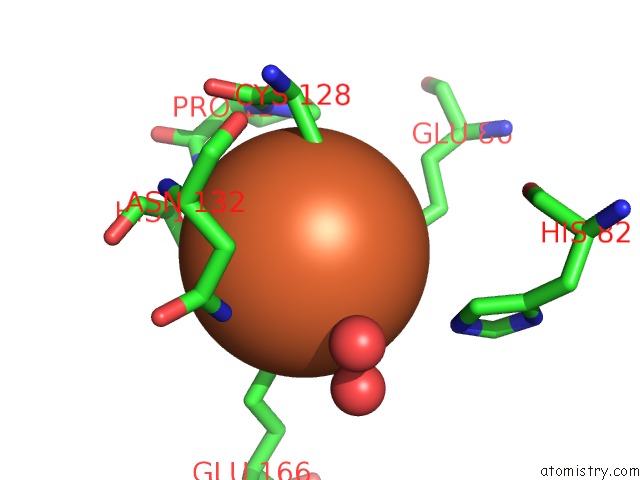
Mono view
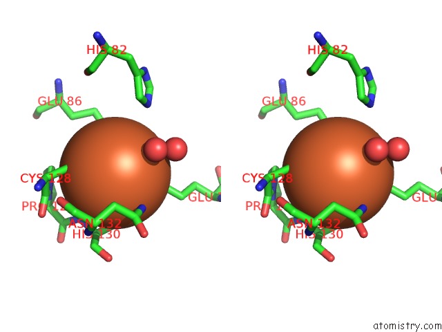
Stereo pair view
Mono view
Stereo pair view
A full contact list of Iron with other atoms in the Fe binding
site number 1 of Crystal Structure of T. Acidophilum Ider within 5.0Å range:
|
Iron binding site 2 out of 2 in 4o5v
Go back to
Iron binding site 2 out
of 2 in the Crystal Structure of T. Acidophilum Ider
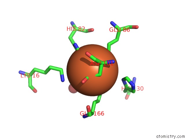
Mono view
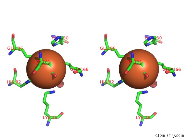
Stereo pair view
Mono view
Stereo pair view
A full contact list of Iron with other atoms in the Fe binding
site number 2 of Crystal Structure of T. Acidophilum Ider within 5.0Å range:
|
Reference:
H.K.Yeo,
Y.W.Park,
J.Y.Lee.
Structural Analysis and Insight Into Metal-Ion Activation of the Iron-Dependent Regulator From Thermoplasma Acidophilum. Acta Crystallogr.,Sect.D V. 70 1281 2014.
ISSN: ISSN 0907-4449
PubMed: 24816097
DOI: 10.1107/S1399004714004118
Page generated: Mon Aug 5 08:06:56 2024
ISSN: ISSN 0907-4449
PubMed: 24816097
DOI: 10.1107/S1399004714004118
Last articles
Zn in 9J0NZn in 9J0O
Zn in 9J0P
Zn in 9FJX
Zn in 9EKB
Zn in 9C0F
Zn in 9CAH
Zn in 9CH0
Zn in 9CH3
Zn in 9CH1