Iron »
PDB 4o1w-4oxf »
4o7g »
Iron in PDB 4o7g: Crystal Structure of Ascorbate-Bound Cytochrome B561, Crystal Soaked in 1 M L-Ascorbate For 40 Minutes
Enzymatic activity of Crystal Structure of Ascorbate-Bound Cytochrome B561, Crystal Soaked in 1 M L-Ascorbate For 40 Minutes
All present enzymatic activity of Crystal Structure of Ascorbate-Bound Cytochrome B561, Crystal Soaked in 1 M L-Ascorbate For 40 Minutes:
1.16.5.1;
1.16.5.1;
Protein crystallography data
The structure of Crystal Structure of Ascorbate-Bound Cytochrome B561, Crystal Soaked in 1 M L-Ascorbate For 40 Minutes, PDB code: 4o7g
was solved by
P.Lu,
D.Ma,
C.Yan,
X.Gong,
M.Du,
Y.Shi,
with X-Ray Crystallography technique. A brief refinement statistics is given in the table below:
| Resolution Low / High (Å) | 28.08 / 2.21 |
| Space group | I 2 2 2 |
| Cell size a, b, c (Å), α, β, γ (°) | 73.186, 108.705, 111.762, 90.00, 90.00, 90.00 |
| R / Rfree (%) | 20.2 / 26.9 |
Iron Binding Sites:
The binding sites of Iron atom in the Crystal Structure of Ascorbate-Bound Cytochrome B561, Crystal Soaked in 1 M L-Ascorbate For 40 Minutes
(pdb code 4o7g). This binding sites where shown within
5.0 Angstroms radius around Iron atom.
In total 4 binding sites of Iron where determined in the Crystal Structure of Ascorbate-Bound Cytochrome B561, Crystal Soaked in 1 M L-Ascorbate For 40 Minutes, PDB code: 4o7g:
Jump to Iron binding site number: 1; 2; 3; 4;
In total 4 binding sites of Iron where determined in the Crystal Structure of Ascorbate-Bound Cytochrome B561, Crystal Soaked in 1 M L-Ascorbate For 40 Minutes, PDB code: 4o7g:
Jump to Iron binding site number: 1; 2; 3; 4;
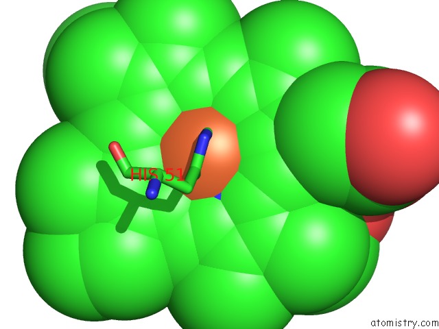
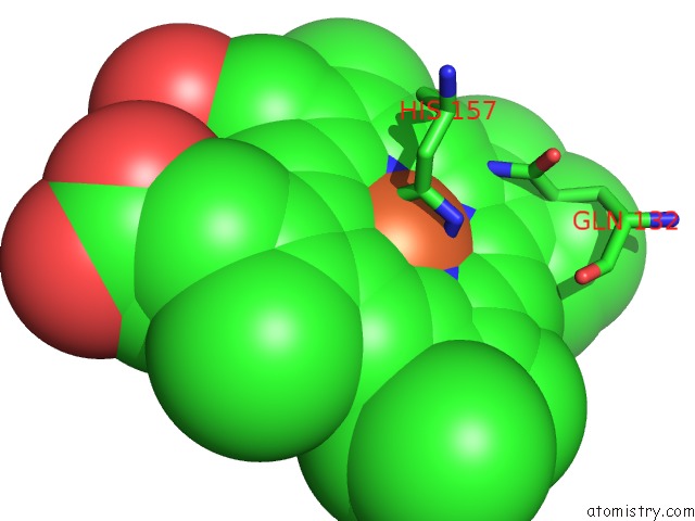
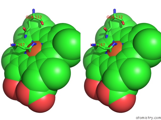
Iron binding site 1 out of 4 in 4o7g
Go back to
Iron binding site 1 out
of 4 in the Crystal Structure of Ascorbate-Bound Cytochrome B561, Crystal Soaked in 1 M L-Ascorbate For 40 Minutes
Mono view

Stereo pair view
Mono view
Stereo pair view
A full contact list of Iron with other atoms in the Fe binding
site number 1 of Crystal Structure of Ascorbate-Bound Cytochrome B561, Crystal Soaked in 1 M L-Ascorbate For 40 Minutes within 5.0Å range:
|
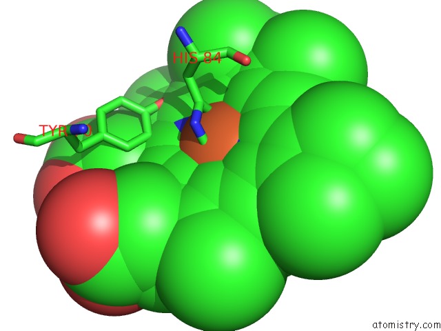
Iron binding site 2 out of 4 in 4o7g
Go back to
Iron binding site 2 out
of 4 in the Crystal Structure of Ascorbate-Bound Cytochrome B561, Crystal Soaked in 1 M L-Ascorbate For 40 Minutes
Mono view
Stereo pair view
Mono view
Stereo pair view
A full contact list of Iron with other atoms in the Fe binding
site number 2 of Crystal Structure of Ascorbate-Bound Cytochrome B561, Crystal Soaked in 1 M L-Ascorbate For 40 Minutes within 5.0Å range:
|
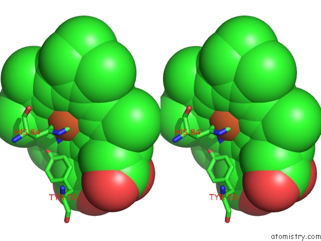
Iron binding site 3 out of 4 in 4o7g
Go back to
Iron binding site 3 out
of 4 in the Crystal Structure of Ascorbate-Bound Cytochrome B561, Crystal Soaked in 1 M L-Ascorbate For 40 Minutes
Mono view

Stereo pair view
Mono view
Stereo pair view
A full contact list of Iron with other atoms in the Fe binding
site number 3 of Crystal Structure of Ascorbate-Bound Cytochrome B561, Crystal Soaked in 1 M L-Ascorbate For 40 Minutes within 5.0Å range:
|
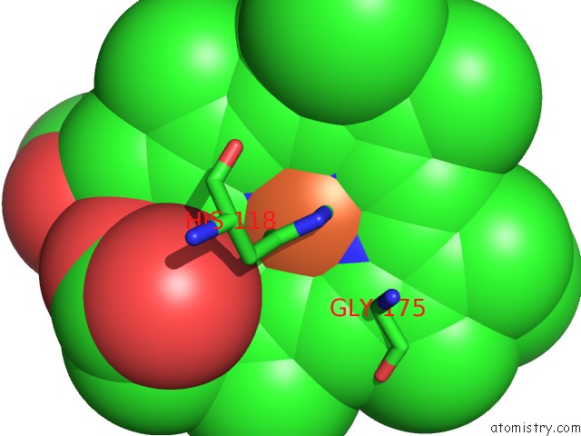
Iron binding site 4 out of 4 in 4o7g
Go back to
Iron binding site 4 out
of 4 in the Crystal Structure of Ascorbate-Bound Cytochrome B561, Crystal Soaked in 1 M L-Ascorbate For 40 Minutes
Mono view
Stereo pair view
Mono view
Stereo pair view
A full contact list of Iron with other atoms in the Fe binding
site number 4 of Crystal Structure of Ascorbate-Bound Cytochrome B561, Crystal Soaked in 1 M L-Ascorbate For 40 Minutes within 5.0Å range:
|
Reference:
P.Lu,
D.Ma,
C.Yan,
X.Gong,
M.Du,
Y.Shi.
Structure and Mechanism of A Eukaryotic Transmembrane Ascorbate-Dependent Oxidoreductase Proc.Natl.Acad.Sci.Usa V. 111 1813 2014.
ISSN: ISSN 0027-8424
PubMed: 24449903
DOI: 10.1073/PNAS.1323931111
Page generated: Mon Aug 5 08:08:32 2024
ISSN: ISSN 0027-8424
PubMed: 24449903
DOI: 10.1073/PNAS.1323931111
Last articles
Zn in 9J0NZn in 9J0O
Zn in 9J0P
Zn in 9FJX
Zn in 9EKB
Zn in 9C0F
Zn in 9CAH
Zn in 9CH0
Zn in 9CH3
Zn in 9CH1