Iron »
PDB 4o1w-4oxf »
4oo7 »
Iron in PDB 4oo7: The 1.55A Crystal Structure of NAF1 (MINER1): the Redox-Active 2FE-2S Protein
Protein crystallography data
The structure of The 1.55A Crystal Structure of NAF1 (MINER1): the Redox-Active 2FE-2S Protein, PDB code: 4oo7
was solved by
S.Tamir,
Y.Eisenberg-Domovich,
A.R.Colman,
J.T.Stofleth,
C.H.Lipper,
M.L.Paddock,
P.A.Jenning,
O.Livnah,
R.Nechushtai,
with X-Ray Crystallography technique. A brief refinement statistics is given in the table below:
| Resolution Low / High (Å) | 31.38 / 1.65 |
| Space group | P 21 21 21 |
| Cell size a, b, c (Å), α, β, γ (°) | 41.009, 48.652, 73.730, 90.00, 90.00, 90.00 |
| R / Rfree (%) | 13.5 / 15 |
Iron Binding Sites:
The binding sites of Iron atom in the The 1.55A Crystal Structure of NAF1 (MINER1): the Redox-Active 2FE-2S Protein
(pdb code 4oo7). This binding sites where shown within
5.0 Angstroms radius around Iron atom.
In total 4 binding sites of Iron where determined in the The 1.55A Crystal Structure of NAF1 (MINER1): the Redox-Active 2FE-2S Protein, PDB code: 4oo7:
Jump to Iron binding site number: 1; 2; 3; 4;
In total 4 binding sites of Iron where determined in the The 1.55A Crystal Structure of NAF1 (MINER1): the Redox-Active 2FE-2S Protein, PDB code: 4oo7:
Jump to Iron binding site number: 1; 2; 3; 4;
Iron binding site 1 out of 4 in 4oo7
Go back to
Iron binding site 1 out
of 4 in the The 1.55A Crystal Structure of NAF1 (MINER1): the Redox-Active 2FE-2S Protein

Mono view
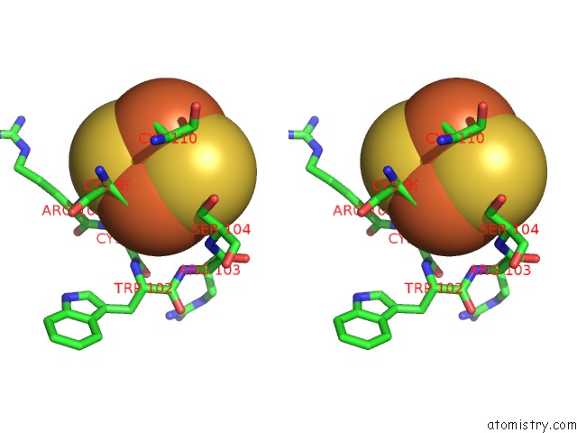
Stereo pair view
Mono view
Stereo pair view
A full contact list of Iron with other atoms in the Fe binding
site number 1 of The 1.55A Crystal Structure of NAF1 (MINER1): the Redox-Active 2FE-2S Protein within 5.0Å range:
|
Iron binding site 2 out of 4 in 4oo7
Go back to
Iron binding site 2 out
of 4 in the The 1.55A Crystal Structure of NAF1 (MINER1): the Redox-Active 2FE-2S Protein
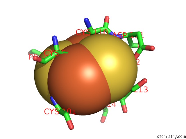
Mono view

Stereo pair view
Mono view
Stereo pair view
A full contact list of Iron with other atoms in the Fe binding
site number 2 of The 1.55A Crystal Structure of NAF1 (MINER1): the Redox-Active 2FE-2S Protein within 5.0Å range:
|
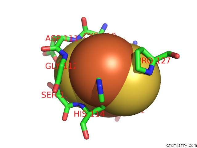
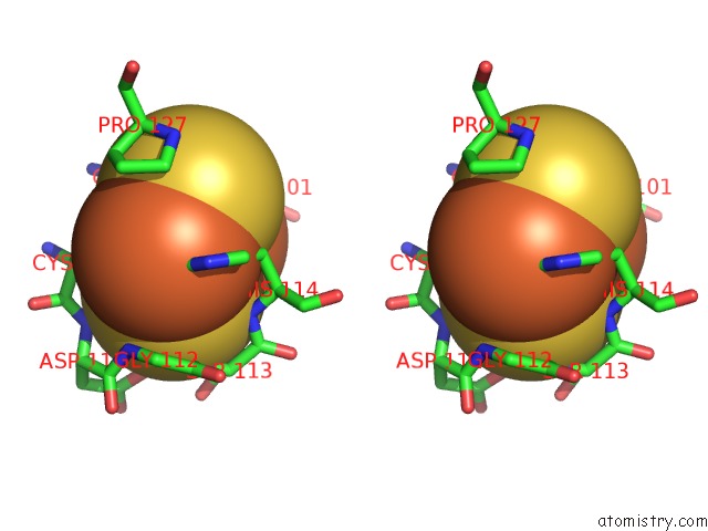
Iron binding site 3 out of 4 in 4oo7
Go back to
Iron binding site 3 out
of 4 in the The 1.55A Crystal Structure of NAF1 (MINER1): the Redox-Active 2FE-2S Protein

Mono view

Stereo pair view
Mono view
Stereo pair view
A full contact list of Iron with other atoms in the Fe binding
site number 3 of The 1.55A Crystal Structure of NAF1 (MINER1): the Redox-Active 2FE-2S Protein within 5.0Å range:
|
Iron binding site 4 out of 4 in 4oo7
Go back to
Iron binding site 4 out
of 4 in the The 1.55A Crystal Structure of NAF1 (MINER1): the Redox-Active 2FE-2S Protein
Mono view
Stereo pair view
Mono view
Stereo pair view
A full contact list of Iron with other atoms in the Fe binding
site number 4 of The 1.55A Crystal Structure of NAF1 (MINER1): the Redox-Active 2FE-2S Protein within 5.0Å range:
|
Reference:
S.Tamir,
Y.Eisenberg-Domovich,
A.R.Conlan,
J.T.Stofleth,
C.H.Lipper,
M.L.Paddock,
R.Mittler,
P.A.Jennings,
O.Livnah,
R.Nechushtai.
A Point Mutation in the [2FE-2S] Cluster Binding Region of the Naf-1 Protein (H114C) Dramatically Hinders the Cluster Donor Properties. Acta Crystallogr.,Sect.D V. 70 1572 2014.
ISSN: ISSN 0907-4449
PubMed: 24914968
DOI: 10.1107/S1399004714005458
Page generated: Mon Aug 5 08:10:18 2024
ISSN: ISSN 0907-4449
PubMed: 24914968
DOI: 10.1107/S1399004714005458
Last articles
Zn in 9J0NZn in 9J0O
Zn in 9J0P
Zn in 9FJX
Zn in 9EKB
Zn in 9C0F
Zn in 9CAH
Zn in 9CH0
Zn in 9CH3
Zn in 9CH1