Iron »
PDB 4o1w-4oxf »
4ou9 »
Iron in PDB 4ou9: Crystal Structure of Apocarotenoid Oxygenase in the Presence of Triton X-100
Enzymatic activity of Crystal Structure of Apocarotenoid Oxygenase in the Presence of Triton X-100
All present enzymatic activity of Crystal Structure of Apocarotenoid Oxygenase in the Presence of Triton X-100:
1.13.11.75;
1.13.11.75;
Protein crystallography data
The structure of Crystal Structure of Apocarotenoid Oxygenase in the Presence of Triton X-100, PDB code: 4ou9
was solved by
X.Sui,
K.Palczewski,
P.D.Kiser,
with X-Ray Crystallography technique. A brief refinement statistics is given in the table below:
| Resolution Low / High (Å) | 48.61 / 2.00 |
| Space group | P 21 21 21 |
| Cell size a, b, c (Å), α, β, γ (°) | 118.470, 125.390, 202.540, 90.00, 90.00, 90.00 |
| R / Rfree (%) | 17.7 / 20.5 |
Other elements in 4ou9:
The structure of Crystal Structure of Apocarotenoid Oxygenase in the Presence of Triton X-100 also contains other interesting chemical elements:
| Chlorine | (Cl) | 2 atoms |
Iron Binding Sites:
The binding sites of Iron atom in the Crystal Structure of Apocarotenoid Oxygenase in the Presence of Triton X-100
(pdb code 4ou9). This binding sites where shown within
5.0 Angstroms radius around Iron atom.
In total 4 binding sites of Iron where determined in the Crystal Structure of Apocarotenoid Oxygenase in the Presence of Triton X-100, PDB code: 4ou9:
Jump to Iron binding site number: 1; 2; 3; 4;
In total 4 binding sites of Iron where determined in the Crystal Structure of Apocarotenoid Oxygenase in the Presence of Triton X-100, PDB code: 4ou9:
Jump to Iron binding site number: 1; 2; 3; 4;
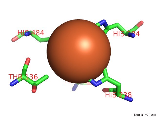
Iron binding site 1 out of 4 in 4ou9
Go back to
Iron binding site 1 out
of 4 in the Crystal Structure of Apocarotenoid Oxygenase in the Presence of Triton X-100
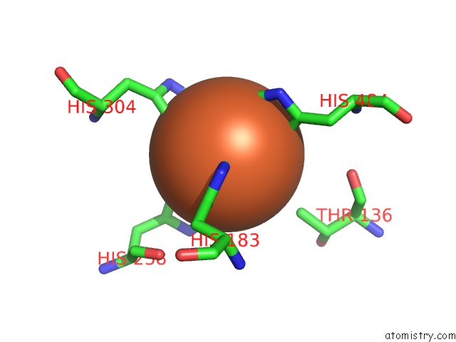
Mono view
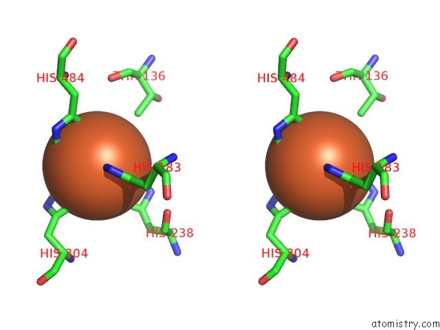
Stereo pair view
Mono view
Stereo pair view
A full contact list of Iron with other atoms in the Fe binding
site number 1 of Crystal Structure of Apocarotenoid Oxygenase in the Presence of Triton X-100 within 5.0Å range:
|
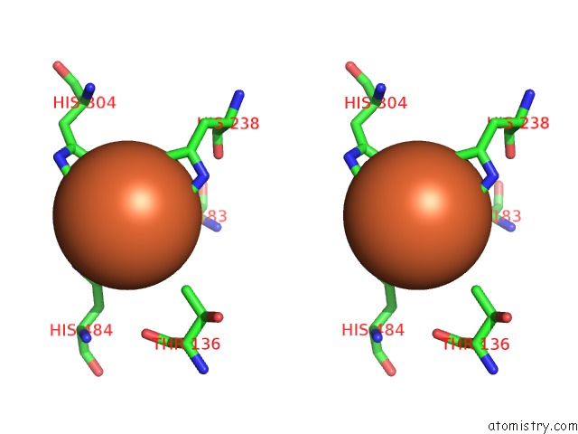
Iron binding site 2 out of 4 in 4ou9
Go back to
Iron binding site 2 out
of 4 in the Crystal Structure of Apocarotenoid Oxygenase in the Presence of Triton X-100
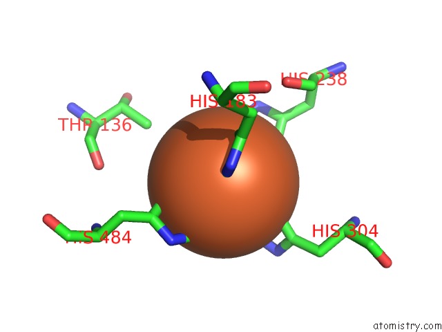
Mono view
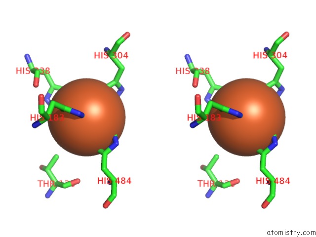
Stereo pair view
Mono view
Stereo pair view
A full contact list of Iron with other atoms in the Fe binding
site number 2 of Crystal Structure of Apocarotenoid Oxygenase in the Presence of Triton X-100 within 5.0Å range:
|
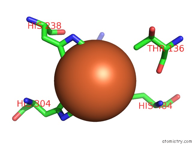
Iron binding site 3 out of 4 in 4ou9
Go back to
Iron binding site 3 out
of 4 in the Crystal Structure of Apocarotenoid Oxygenase in the Presence of Triton X-100
Mono view
Stereo pair view
Mono view
Stereo pair view
A full contact list of Iron with other atoms in the Fe binding
site number 3 of Crystal Structure of Apocarotenoid Oxygenase in the Presence of Triton X-100 within 5.0Å range:
|
Iron binding site 4 out of 4 in 4ou9
Go back to
Iron binding site 4 out
of 4 in the Crystal Structure of Apocarotenoid Oxygenase in the Presence of Triton X-100
Mono view
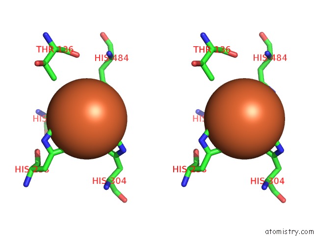
Stereo pair view
Mono view
Stereo pair view
A full contact list of Iron with other atoms in the Fe binding
site number 4 of Crystal Structure of Apocarotenoid Oxygenase in the Presence of Triton X-100 within 5.0Å range:
|
Reference:
X.Sui,
P.D.Kiser,
T.Che,
P.R.Carey,
M.Golczak,
W.Shi,
J.Von Lintig,
K.Palczewski.
Analysis of Carotenoid Isomerase Activity in A Prototypical Carotenoid Cleavage Enzyme, Apocarotenoid Oxygenase (Aco). J.Biol.Chem. V. 289 12286 2014.
ISSN: ISSN 0021-9258
PubMed: 24648526
DOI: 10.1074/JBC.M114.552836
Page generated: Tue Aug 5 13:34:40 2025
ISSN: ISSN 0021-9258
PubMed: 24648526
DOI: 10.1074/JBC.M114.552836
Last articles
Fe in 4TODFe in 4TO9
Fe in 4TOB
Fe in 4TKV
Fe in 4TKU
Fe in 4TNK
Fe in 4TNJ
Fe in 4TNI
Fe in 4TNH
Fe in 4TLF