Iron »
PDB 4oyn-4q1o »
4pk6 »
Iron in PDB 4pk6: Crystal Structure of the Indoleamine 2,3-Dioxygenagse 1 (IDO1) Complexed with Imidazothiazole Derivative
Enzymatic activity of Crystal Structure of the Indoleamine 2,3-Dioxygenagse 1 (IDO1) Complexed with Imidazothiazole Derivative
All present enzymatic activity of Crystal Structure of the Indoleamine 2,3-Dioxygenagse 1 (IDO1) Complexed with Imidazothiazole Derivative:
1.13.11.52;
1.13.11.52;
Protein crystallography data
The structure of Crystal Structure of the Indoleamine 2,3-Dioxygenagse 1 (IDO1) Complexed with Imidazothiazole Derivative, PDB code: 4pk6
was solved by
T.Kohno,
S.Tojo,
T.Ishii,
S.Kamioka,
with X-Ray Crystallography technique. A brief refinement statistics is given in the table below:
| Resolution Low / High (Å) | 75.65 / 3.45 |
| Space group | P 21 21 21 |
| Cell size a, b, c (Å), α, β, γ (°) | 84.080, 91.120, 135.690, 90.00, 90.00, 90.00 |
| R / Rfree (%) | 19 / 25.6 |
Other elements in 4pk6:
The structure of Crystal Structure of the Indoleamine 2,3-Dioxygenagse 1 (IDO1) Complexed with Imidazothiazole Derivative also contains other interesting chemical elements:
| Chlorine | (Cl) | 2 atoms |
Iron Binding Sites:
The binding sites of Iron atom in the Crystal Structure of the Indoleamine 2,3-Dioxygenagse 1 (IDO1) Complexed with Imidazothiazole Derivative
(pdb code 4pk6). This binding sites where shown within
5.0 Angstroms radius around Iron atom.
In total 2 binding sites of Iron where determined in the Crystal Structure of the Indoleamine 2,3-Dioxygenagse 1 (IDO1) Complexed with Imidazothiazole Derivative, PDB code: 4pk6:
Jump to Iron binding site number: 1; 2;
In total 2 binding sites of Iron where determined in the Crystal Structure of the Indoleamine 2,3-Dioxygenagse 1 (IDO1) Complexed with Imidazothiazole Derivative, PDB code: 4pk6:
Jump to Iron binding site number: 1; 2;
Iron binding site 1 out of 2 in 4pk6
Go back to
Iron binding site 1 out
of 2 in the Crystal Structure of the Indoleamine 2,3-Dioxygenagse 1 (IDO1) Complexed with Imidazothiazole Derivative
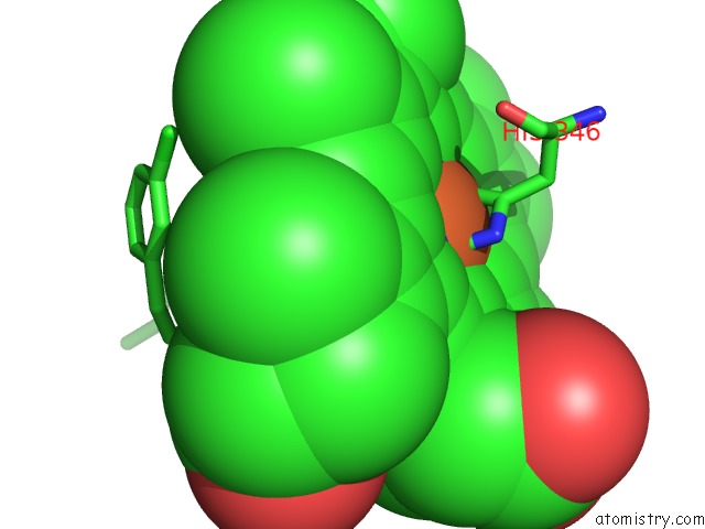
Mono view
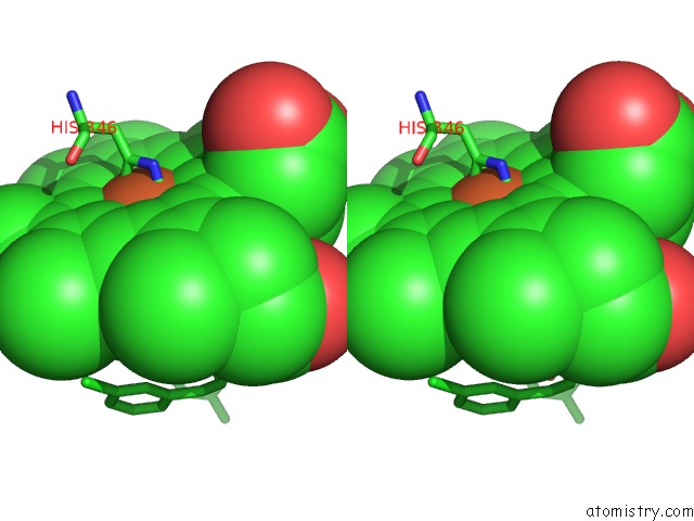
Stereo pair view
Mono view
Stereo pair view
A full contact list of Iron with other atoms in the Fe binding
site number 1 of Crystal Structure of the Indoleamine 2,3-Dioxygenagse 1 (IDO1) Complexed with Imidazothiazole Derivative within 5.0Å range:
|
Iron binding site 2 out of 2 in 4pk6
Go back to
Iron binding site 2 out
of 2 in the Crystal Structure of the Indoleamine 2,3-Dioxygenagse 1 (IDO1) Complexed with Imidazothiazole Derivative

Mono view
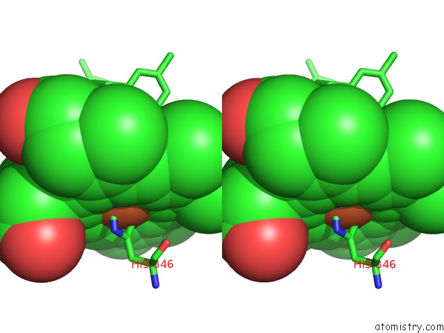
Stereo pair view
Mono view
Stereo pair view
A full contact list of Iron with other atoms in the Fe binding
site number 2 of Crystal Structure of the Indoleamine 2,3-Dioxygenagse 1 (IDO1) Complexed with Imidazothiazole Derivative within 5.0Å range:
|
Reference:
S.Tojo,
T.Kohno,
T.Tanaka,
S.Kamioka,
Y.Ota,
T.Ishii,
K.Kamimoto,
S.Asano,
Y.Isobe.
Crystal Structures and Structure-Activity Relationships of Imidazothiazole Derivatives As IDO1 Inhibitors. Acs Med.Chem.Lett. V. 5 1119 2014.
ISSN: ISSN 1948-5875
PubMed: 25313323
DOI: 10.1021/ML500247W
Page generated: Mon Aug 5 08:28:19 2024
ISSN: ISSN 1948-5875
PubMed: 25313323
DOI: 10.1021/ML500247W
Last articles
Zn in 9MJ5Zn in 9HNW
Zn in 9G0L
Zn in 9FNE
Zn in 9DZN
Zn in 9E0I
Zn in 9D32
Zn in 9DAK
Zn in 8ZXC
Zn in 8ZUF