Iron »
PDB 4oyn-4q1o »
4pl1 »
Iron in PDB 4pl1: X-Ray Crystal Structure of C118A Rlmn From Escherichia Coli with S- Adenosylmethionine
Enzymatic activity of X-Ray Crystal Structure of C118A Rlmn From Escherichia Coli with S- Adenosylmethionine
All present enzymatic activity of X-Ray Crystal Structure of C118A Rlmn From Escherichia Coli with S- Adenosylmethionine:
2.1.1.192;
2.1.1.192;
Protein crystallography data
The structure of X-Ray Crystal Structure of C118A Rlmn From Escherichia Coli with S- Adenosylmethionine, PDB code: 4pl1
was solved by
A.K.Boal,
A.C.Rosenzweig,
with X-Ray Crystallography technique. A brief refinement statistics is given in the table below:
| Resolution Low / High (Å) | 28.85 / 2.58 |
| Space group | P 21 21 21 |
| Cell size a, b, c (Å), α, β, γ (°) | 55.627, 55.729, 254.032, 90.00, 90.00, 90.00 |
| R / Rfree (%) | 26.3 / 30 |
Iron Binding Sites:
The binding sites of Iron atom in the X-Ray Crystal Structure of C118A Rlmn From Escherichia Coli with S- Adenosylmethionine
(pdb code 4pl1). This binding sites where shown within
5.0 Angstroms radius around Iron atom.
In total 8 binding sites of Iron where determined in the X-Ray Crystal Structure of C118A Rlmn From Escherichia Coli with S- Adenosylmethionine, PDB code: 4pl1:
Jump to Iron binding site number: 1; 2; 3; 4; 5; 6; 7; 8;
In total 8 binding sites of Iron where determined in the X-Ray Crystal Structure of C118A Rlmn From Escherichia Coli with S- Adenosylmethionine, PDB code: 4pl1:
Jump to Iron binding site number: 1; 2; 3; 4; 5; 6; 7; 8;
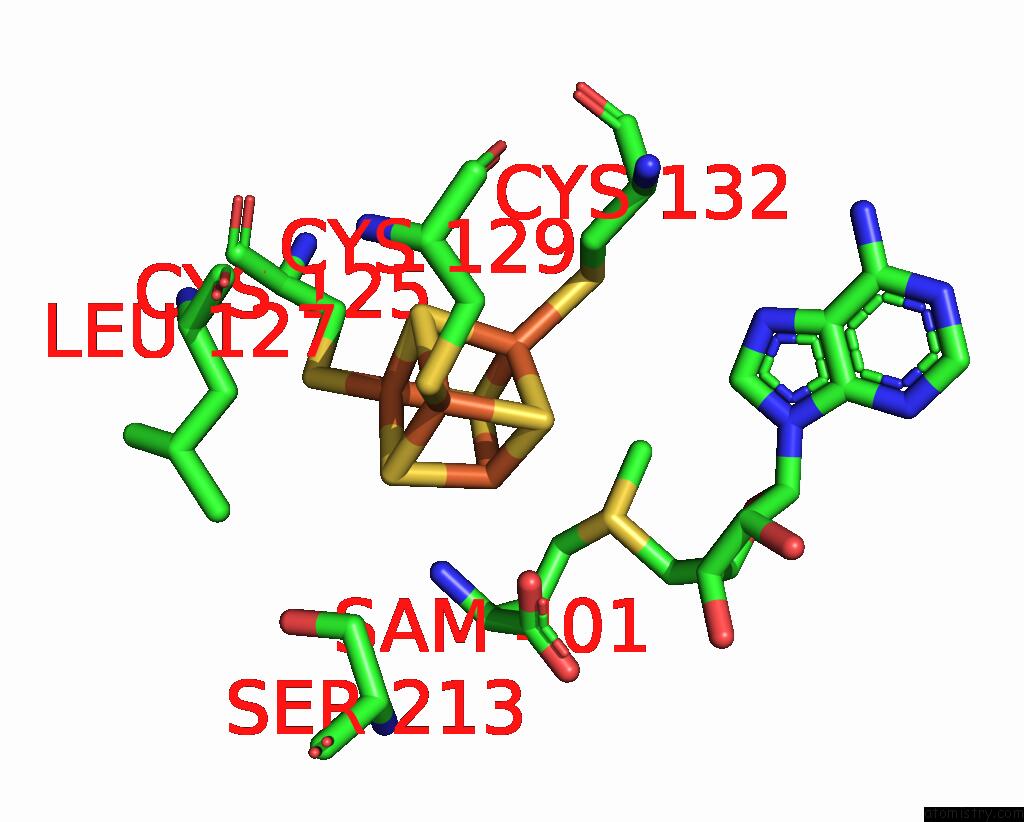
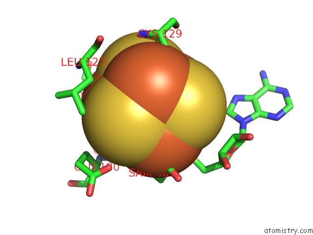
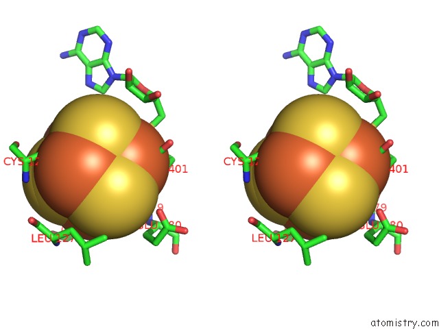
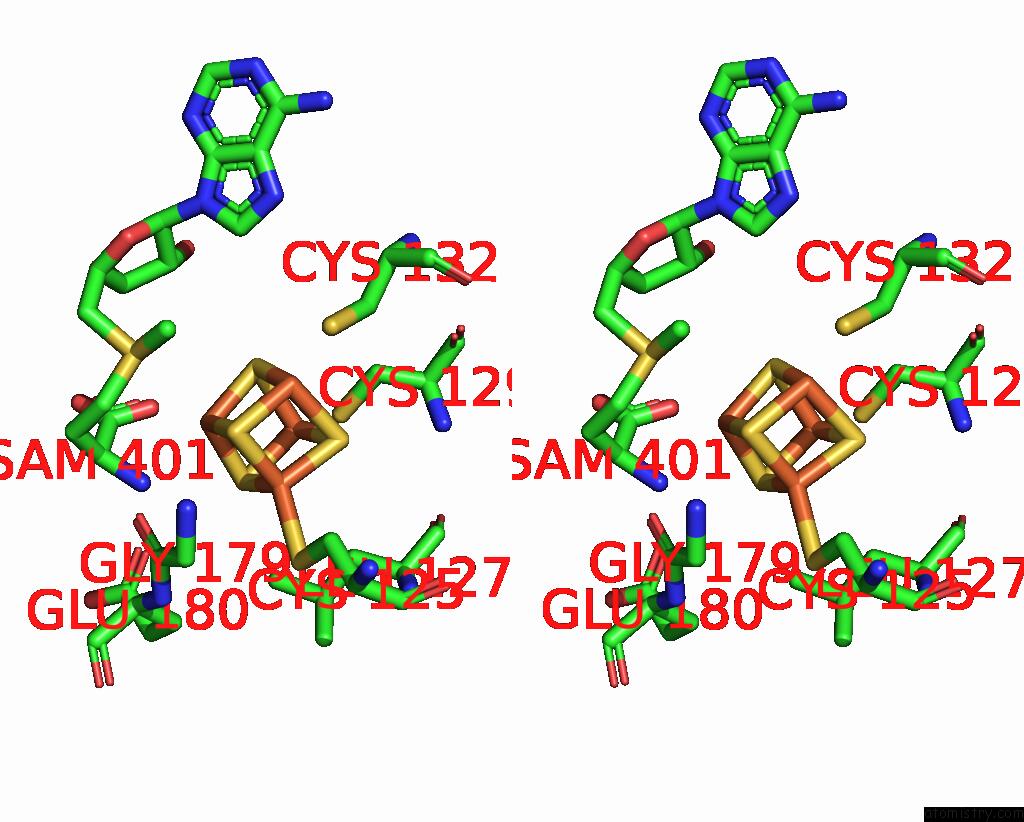
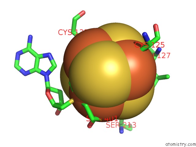
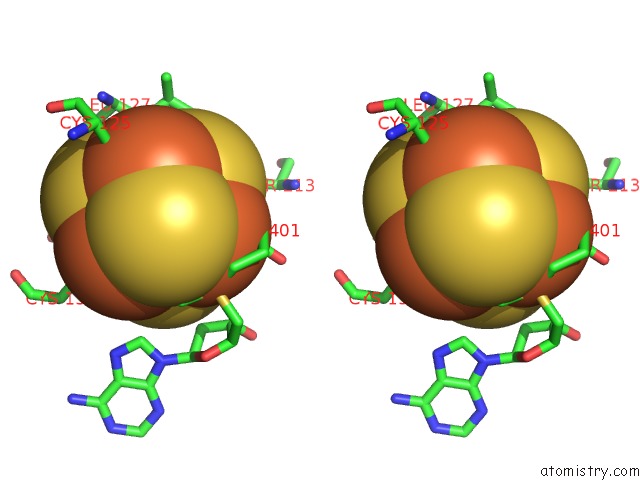
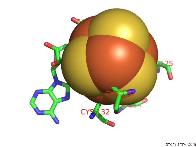
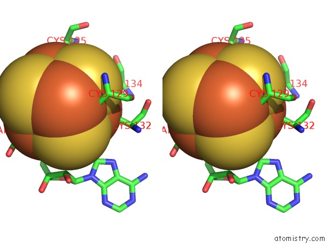
Iron binding site 1 out of 8 in 4pl1
Go back to
Iron binding site 1 out
of 8 in the X-Ray Crystal Structure of C118A Rlmn From Escherichia Coli with S- Adenosylmethionine
Mono view
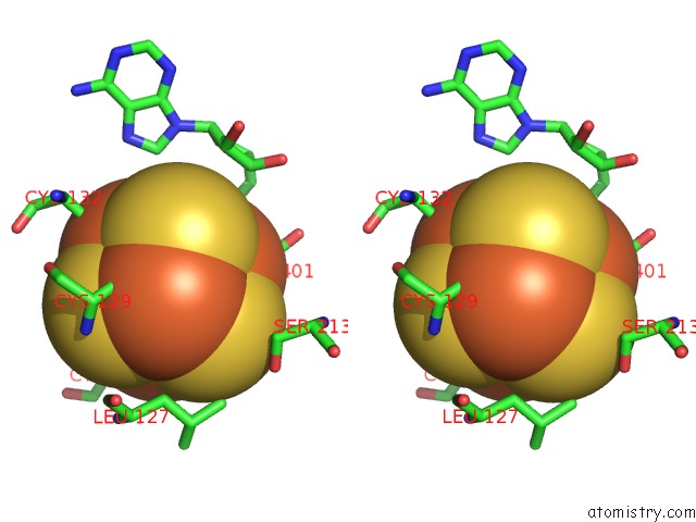
Stereo pair view
Mono view
Stereo pair view
A full contact list of Iron with other atoms in the Fe binding
site number 1 of X-Ray Crystal Structure of C118A Rlmn From Escherichia Coli with S- Adenosylmethionine within 5.0Å range:
|
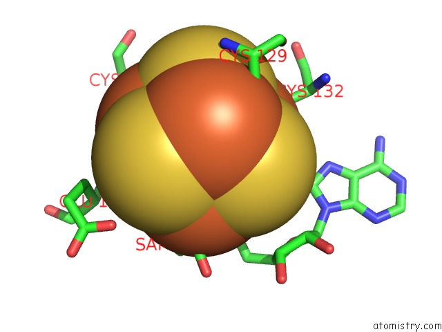
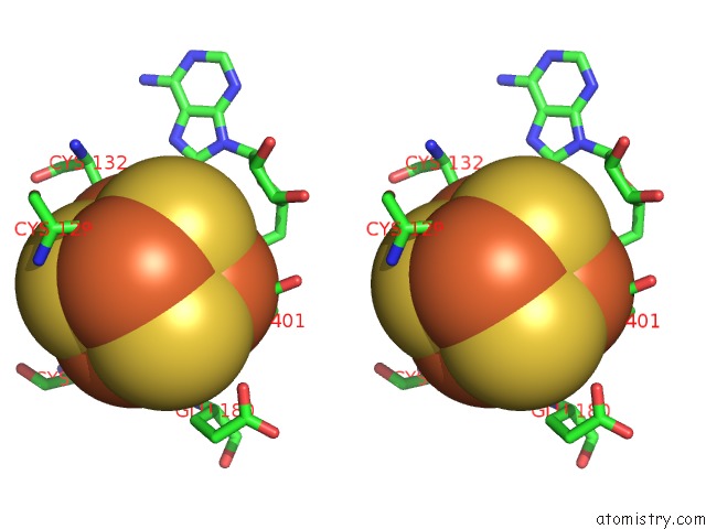
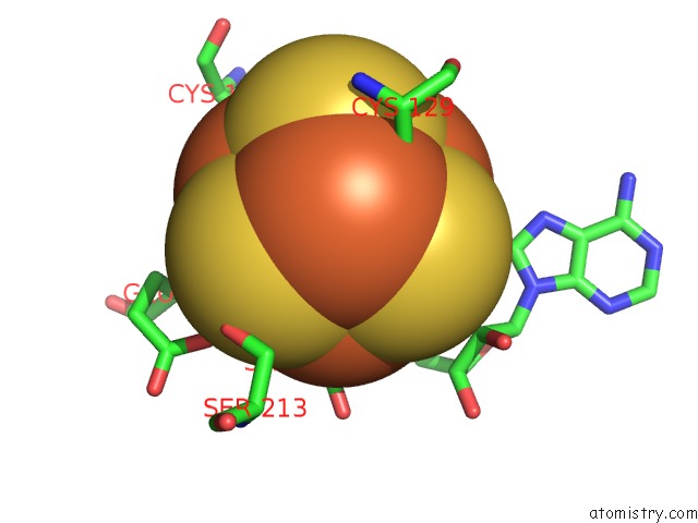
Iron binding site 2 out of 8 in 4pl1
Go back to
Iron binding site 2 out
of 8 in the X-Ray Crystal Structure of C118A Rlmn From Escherichia Coli with S- Adenosylmethionine
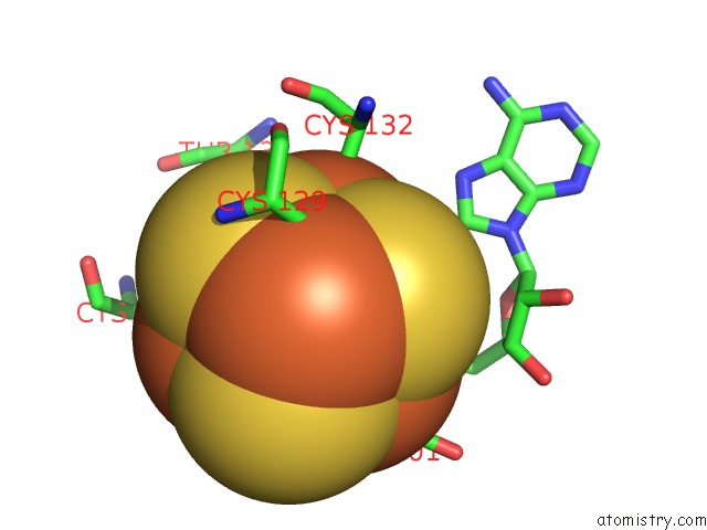
Mono view
Stereo pair view
Mono view
Stereo pair view
A full contact list of Iron with other atoms in the Fe binding
site number 2 of X-Ray Crystal Structure of C118A Rlmn From Escherichia Coli with S- Adenosylmethionine within 5.0Å range:
|
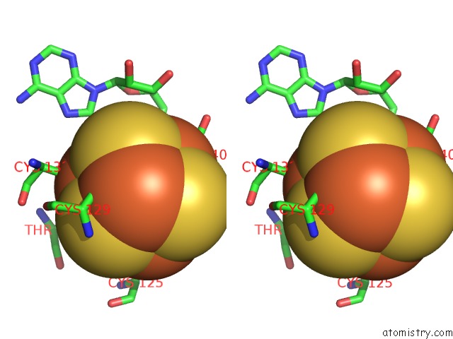
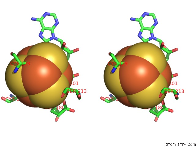
Iron binding site 3 out of 8 in 4pl1
Go back to
Iron binding site 3 out
of 8 in the X-Ray Crystal Structure of C118A Rlmn From Escherichia Coli with S- Adenosylmethionine
Mono view
Stereo pair view
Mono view
Stereo pair view
A full contact list of Iron with other atoms in the Fe binding
site number 3 of X-Ray Crystal Structure of C118A Rlmn From Escherichia Coli with S- Adenosylmethionine within 5.0Å range:
|
Iron binding site 4 out of 8 in 4pl1
Go back to
Iron binding site 4 out
of 8 in the X-Ray Crystal Structure of C118A Rlmn From Escherichia Coli with S- Adenosylmethionine
Mono view
Stereo pair view
Mono view
Stereo pair view
A full contact list of Iron with other atoms in the Fe binding
site number 4 of X-Ray Crystal Structure of C118A Rlmn From Escherichia Coli with S- Adenosylmethionine within 5.0Å range:
|
Iron binding site 5 out of 8 in 4pl1
Go back to
Iron binding site 5 out
of 8 in the X-Ray Crystal Structure of C118A Rlmn From Escherichia Coli with S- Adenosylmethionine

Mono view
Stereo pair view
Mono view
Stereo pair view
A full contact list of Iron with other atoms in the Fe binding
site number 5 of X-Ray Crystal Structure of C118A Rlmn From Escherichia Coli with S- Adenosylmethionine within 5.0Å range:
|
Iron binding site 6 out of 8 in 4pl1
Go back to
Iron binding site 6 out
of 8 in the X-Ray Crystal Structure of C118A Rlmn From Escherichia Coli with S- Adenosylmethionine
Mono view
Stereo pair view
Mono view
Stereo pair view
A full contact list of Iron with other atoms in the Fe binding
site number 6 of X-Ray Crystal Structure of C118A Rlmn From Escherichia Coli with S- Adenosylmethionine within 5.0Å range:
|
Iron binding site 7 out of 8 in 4pl1
Go back to
Iron binding site 7 out
of 8 in the X-Ray Crystal Structure of C118A Rlmn From Escherichia Coli with S- Adenosylmethionine
Mono view
Stereo pair view
Mono view
Stereo pair view
A full contact list of Iron with other atoms in the Fe binding
site number 7 of X-Ray Crystal Structure of C118A Rlmn From Escherichia Coli with S- Adenosylmethionine within 5.0Å range:
|
Iron binding site 8 out of 8 in 4pl1
Go back to
Iron binding site 8 out
of 8 in the X-Ray Crystal Structure of C118A Rlmn From Escherichia Coli with S- Adenosylmethionine
Mono view
Stereo pair view
Mono view
Stereo pair view
A full contact list of Iron with other atoms in the Fe binding
site number 8 of X-Ray Crystal Structure of C118A Rlmn From Escherichia Coli with S- Adenosylmethionine within 5.0Å range:
|
Reference:
A.Silakov,
T.L.Grove,
M.I.Radle,
M.R.Bauerle,
M.T.Green,
A.C.Rosenzweig,
A.K.Boal,
S.J.Booker.
Characterization of A Cross-Linked Protein-Nucleic Acid Substrate Radical in the Reaction Catalyzed By Rlmn. J.Am.Chem.Soc. V. 136 8221 2014.
ISSN: ESSN 1520-5126
PubMed: 24806349
DOI: 10.1021/JA410560P
Page generated: Mon Aug 5 08:28:35 2024
ISSN: ESSN 1520-5126
PubMed: 24806349
DOI: 10.1021/JA410560P
Last articles
Fe in 2YXOFe in 2YRS
Fe in 2YXC
Fe in 2YNM
Fe in 2YVJ
Fe in 2YP1
Fe in 2YU2
Fe in 2YU1
Fe in 2YQB
Fe in 2YOO