Iron »
PDB 4r33-4rxn »
4rb4 »
Iron in PDB 4rb4: Crystal Structure of Dodecameric Iron-Containing Heptosyltransferase Tibc in Complex with Adp-D-Beta-D-Heptose at 3.9 Angstrom Resolution
Protein crystallography data
The structure of Crystal Structure of Dodecameric Iron-Containing Heptosyltransferase Tibc in Complex with Adp-D-Beta-D-Heptose at 3.9 Angstrom Resolution, PDB code: 4rb4
was solved by
Q.Yao,
Q.Lu,
F.Shao,
with X-Ray Crystallography technique. A brief refinement statistics is given in the table below:
| Resolution Low / High (Å) | 20.04 / 3.88 |
| Space group | P 1 21 1 |
| Cell size a, b, c (Å), α, β, γ (°) | 88.292, 313.196, 164.695, 90.00, 101.26, 90.00 |
| R / Rfree (%) | 28.2 / 29.5 |
Iron Binding Sites:
Pages:
>>> Page 1 <<< Page 2, Binding sites: 11 - 12;Binding sites:
The binding sites of Iron atom in the Crystal Structure of Dodecameric Iron-Containing Heptosyltransferase Tibc in Complex with Adp-D-Beta-D-Heptose at 3.9 Angstrom Resolution (pdb code 4rb4). This binding sites where shown within 5.0 Angstroms radius around Iron atom.In total 12 binding sites of Iron where determined in the Crystal Structure of Dodecameric Iron-Containing Heptosyltransferase Tibc in Complex with Adp-D-Beta-D-Heptose at 3.9 Angstrom Resolution, PDB code: 4rb4:
Jump to Iron binding site number: 1; 2; 3; 4; 5; 6; 7; 8; 9; 10;
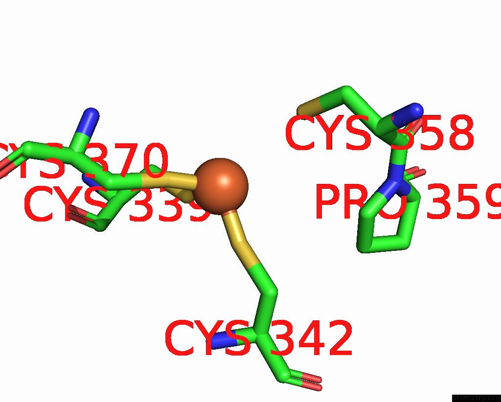
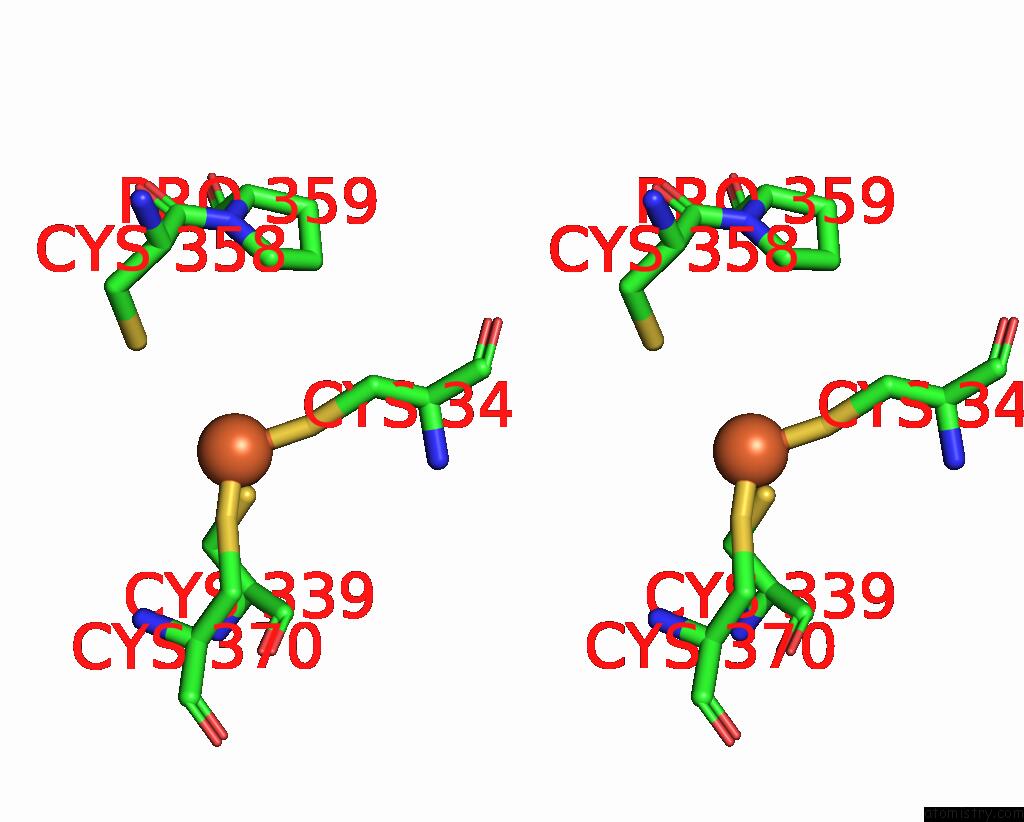
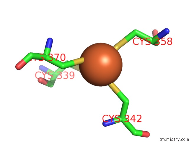
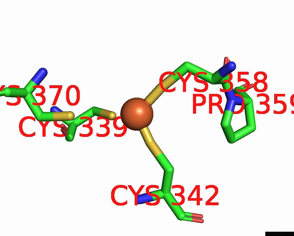
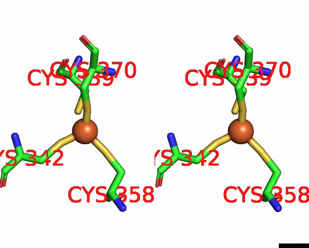
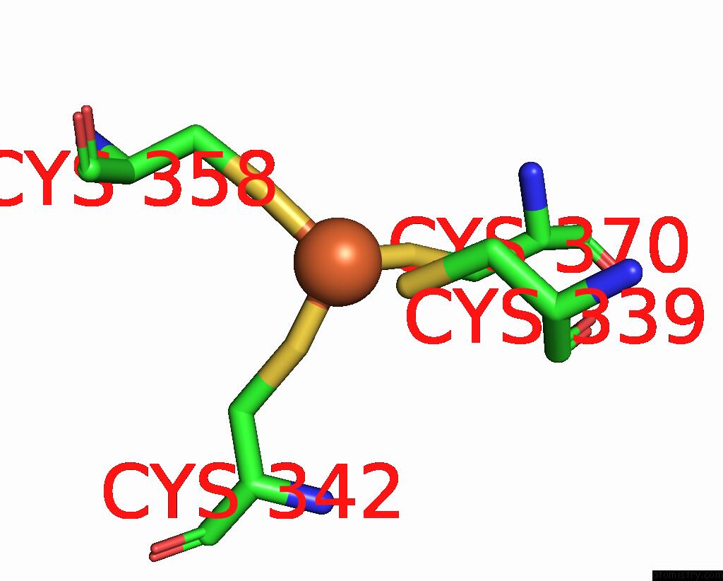
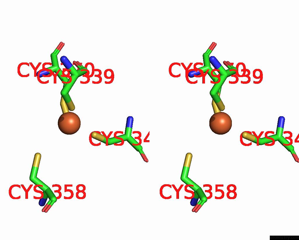
Iron binding site 1 out of 12 in 4rb4
Go back to
Iron binding site 1 out
of 12 in the Crystal Structure of Dodecameric Iron-Containing Heptosyltransferase Tibc in Complex with Adp-D-Beta-D-Heptose at 3.9 Angstrom Resolution
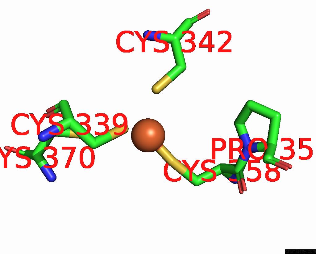
Mono view
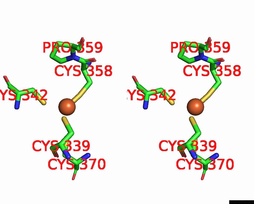
Stereo pair view
Mono view
Stereo pair view
A full contact list of Iron with other atoms in the Fe binding
site number 1 of Crystal Structure of Dodecameric Iron-Containing Heptosyltransferase Tibc in Complex with Adp-D-Beta-D-Heptose at 3.9 Angstrom Resolution within 5.0Å range:
|
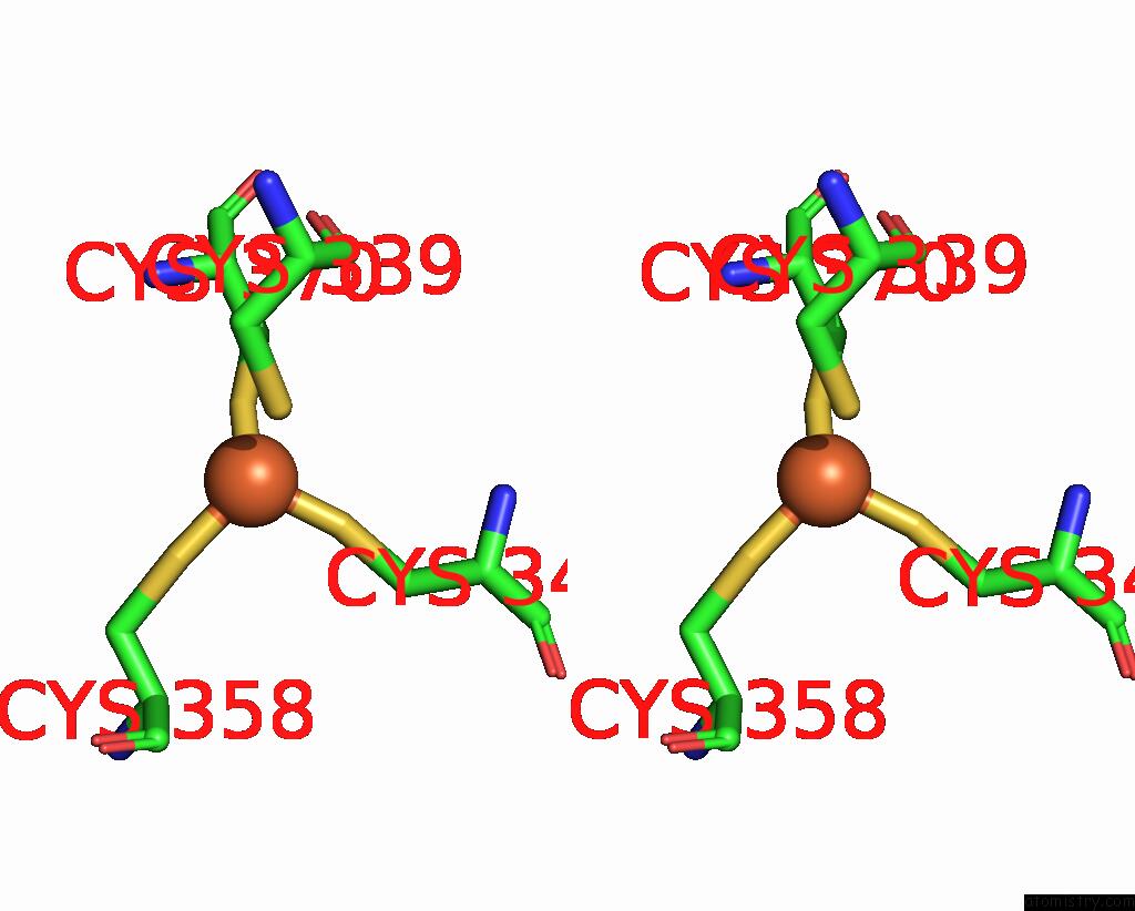
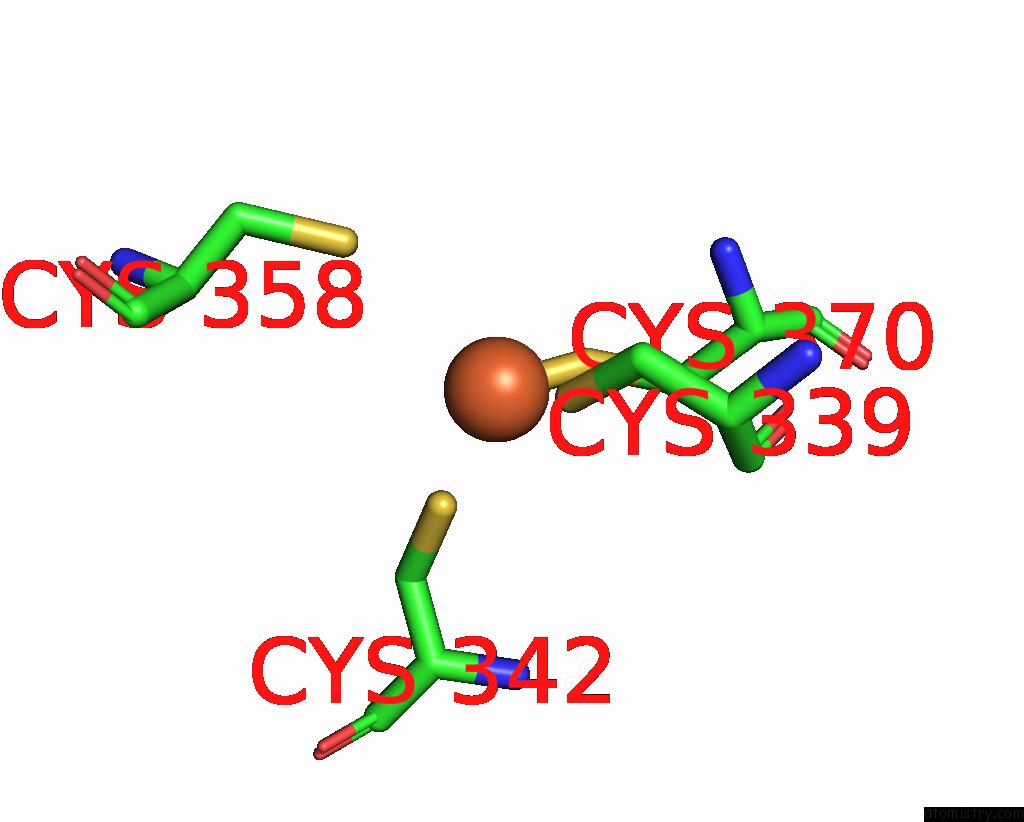
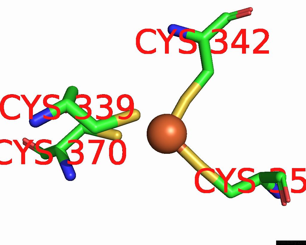
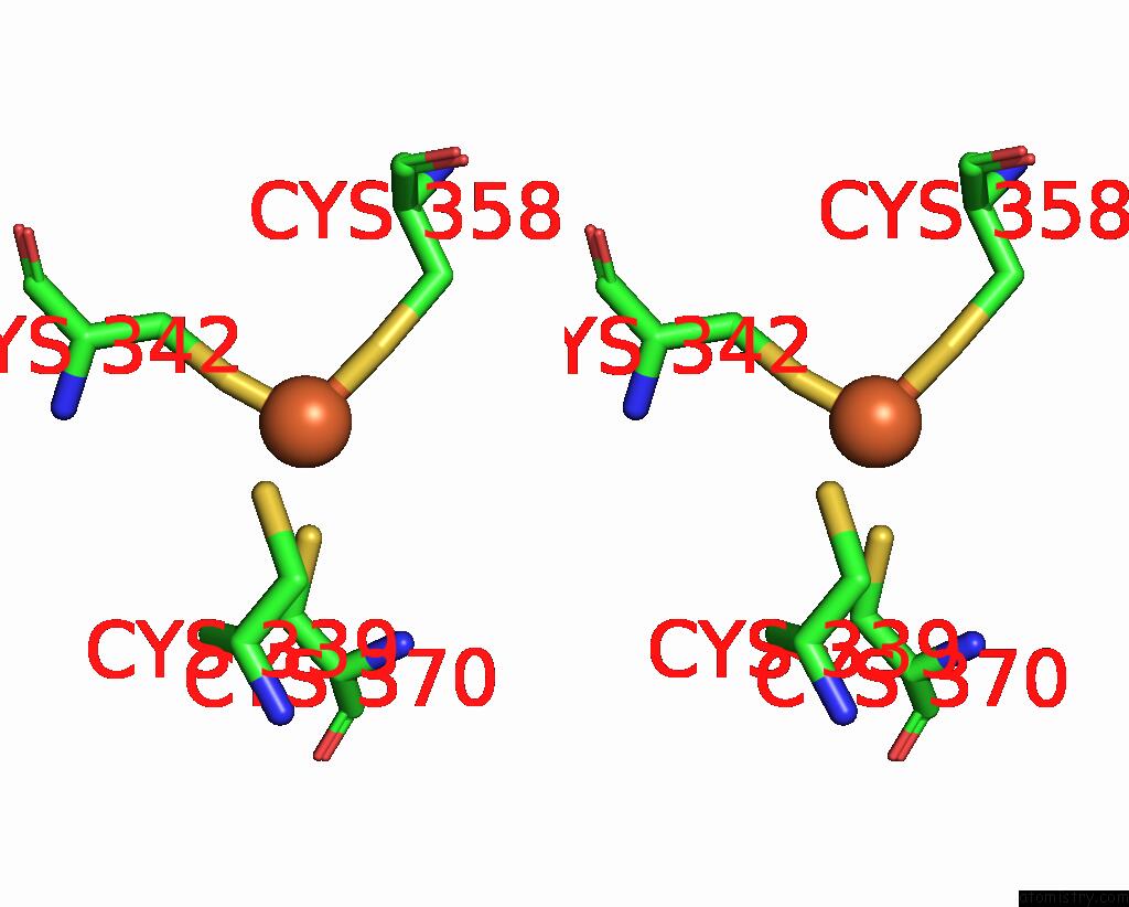
Iron binding site 2 out of 12 in 4rb4
Go back to
Iron binding site 2 out
of 12 in the Crystal Structure of Dodecameric Iron-Containing Heptosyltransferase Tibc in Complex with Adp-D-Beta-D-Heptose at 3.9 Angstrom Resolution
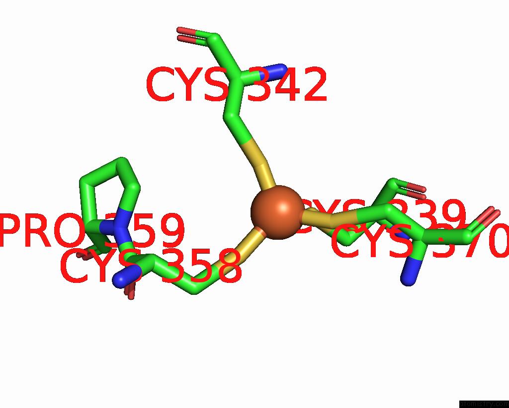
Mono view
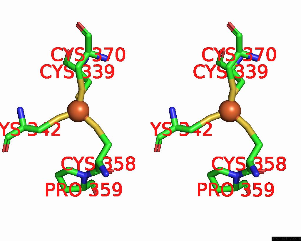
Stereo pair view
Mono view
Stereo pair view
A full contact list of Iron with other atoms in the Fe binding
site number 2 of Crystal Structure of Dodecameric Iron-Containing Heptosyltransferase Tibc in Complex with Adp-D-Beta-D-Heptose at 3.9 Angstrom Resolution within 5.0Å range:
|
Iron binding site 3 out of 12 in 4rb4
Go back to
Iron binding site 3 out
of 12 in the Crystal Structure of Dodecameric Iron-Containing Heptosyltransferase Tibc in Complex with Adp-D-Beta-D-Heptose at 3.9 Angstrom Resolution
Mono view
Stereo pair view
Mono view
Stereo pair view
A full contact list of Iron with other atoms in the Fe binding
site number 3 of Crystal Structure of Dodecameric Iron-Containing Heptosyltransferase Tibc in Complex with Adp-D-Beta-D-Heptose at 3.9 Angstrom Resolution within 5.0Å range:
|
Iron binding site 4 out of 12 in 4rb4
Go back to
Iron binding site 4 out
of 12 in the Crystal Structure of Dodecameric Iron-Containing Heptosyltransferase Tibc in Complex with Adp-D-Beta-D-Heptose at 3.9 Angstrom Resolution
Mono view

Stereo pair view
Mono view
Stereo pair view
A full contact list of Iron with other atoms in the Fe binding
site number 4 of Crystal Structure of Dodecameric Iron-Containing Heptosyltransferase Tibc in Complex with Adp-D-Beta-D-Heptose at 3.9 Angstrom Resolution within 5.0Å range:
|
Iron binding site 5 out of 12 in 4rb4
Go back to
Iron binding site 5 out
of 12 in the Crystal Structure of Dodecameric Iron-Containing Heptosyltransferase Tibc in Complex with Adp-D-Beta-D-Heptose at 3.9 Angstrom Resolution
Mono view

Stereo pair view
Mono view
Stereo pair view
A full contact list of Iron with other atoms in the Fe binding
site number 5 of Crystal Structure of Dodecameric Iron-Containing Heptosyltransferase Tibc in Complex with Adp-D-Beta-D-Heptose at 3.9 Angstrom Resolution within 5.0Å range:
|
Iron binding site 6 out of 12 in 4rb4
Go back to
Iron binding site 6 out
of 12 in the Crystal Structure of Dodecameric Iron-Containing Heptosyltransferase Tibc in Complex with Adp-D-Beta-D-Heptose at 3.9 Angstrom Resolution

Mono view
Stereo pair view
Mono view
Stereo pair view
A full contact list of Iron with other atoms in the Fe binding
site number 6 of Crystal Structure of Dodecameric Iron-Containing Heptosyltransferase Tibc in Complex with Adp-D-Beta-D-Heptose at 3.9 Angstrom Resolution within 5.0Å range:
|
Iron binding site 7 out of 12 in 4rb4
Go back to
Iron binding site 7 out
of 12 in the Crystal Structure of Dodecameric Iron-Containing Heptosyltransferase Tibc in Complex with Adp-D-Beta-D-Heptose at 3.9 Angstrom Resolution

Mono view

Stereo pair view
Mono view
Stereo pair view
A full contact list of Iron with other atoms in the Fe binding
site number 7 of Crystal Structure of Dodecameric Iron-Containing Heptosyltransferase Tibc in Complex with Adp-D-Beta-D-Heptose at 3.9 Angstrom Resolution within 5.0Å range:
|
Iron binding site 8 out of 12 in 4rb4
Go back to
Iron binding site 8 out
of 12 in the Crystal Structure of Dodecameric Iron-Containing Heptosyltransferase Tibc in Complex with Adp-D-Beta-D-Heptose at 3.9 Angstrom Resolution
Mono view
Stereo pair view
Mono view
Stereo pair view
A full contact list of Iron with other atoms in the Fe binding
site number 8 of Crystal Structure of Dodecameric Iron-Containing Heptosyltransferase Tibc in Complex with Adp-D-Beta-D-Heptose at 3.9 Angstrom Resolution within 5.0Å range:
|
Iron binding site 9 out of 12 in 4rb4
Go back to
Iron binding site 9 out
of 12 in the Crystal Structure of Dodecameric Iron-Containing Heptosyltransferase Tibc in Complex with Adp-D-Beta-D-Heptose at 3.9 Angstrom Resolution
Mono view
Stereo pair view
Mono view
Stereo pair view
A full contact list of Iron with other atoms in the Fe binding
site number 9 of Crystal Structure of Dodecameric Iron-Containing Heptosyltransferase Tibc in Complex with Adp-D-Beta-D-Heptose at 3.9 Angstrom Resolution within 5.0Å range:
|
Iron binding site 10 out of 12 in 4rb4
Go back to
Iron binding site 10 out
of 12 in the Crystal Structure of Dodecameric Iron-Containing Heptosyltransferase Tibc in Complex with Adp-D-Beta-D-Heptose at 3.9 Angstrom Resolution
Mono view
Stereo pair view
Mono view
Stereo pair view
A full contact list of Iron with other atoms in the Fe binding
site number 10 of Crystal Structure of Dodecameric Iron-Containing Heptosyltransferase Tibc in Complex with Adp-D-Beta-D-Heptose at 3.9 Angstrom Resolution within 5.0Å range:
|
Reference:
Q.Yao,
Q.Lu,
X.Wan,
F.Song,
Y.Xu,
M.Hu,
A.Zamyatina,
X.Liu,
N.Huang,
P.Zhu,
F.Shao.
A Structural Mechanism For Bacterial Autotransporter Glycosylation By A Dodecameric Heptosyltransferase Family Elife V. 3 2014.
ISSN: ESSN 2050-084X
PubMed: 25310236
DOI: 10.7554/ELIFE.03714
Page generated: Mon Aug 5 09:10:00 2024
ISSN: ESSN 2050-084X
PubMed: 25310236
DOI: 10.7554/ELIFE.03714
Last articles
Zn in 9J0NZn in 9J0O
Zn in 9J0P
Zn in 9FJX
Zn in 9EKB
Zn in 9C0F
Zn in 9CAH
Zn in 9CH0
Zn in 9CH3
Zn in 9CH1