Iron »
PDB 4r33-4rxn »
4rxn »
Iron in PDB 4rxn: Crystallographic Refinement of Rubredoxin at 1.2 Angstroms Resolution
Protein crystallography data
The structure of Crystallographic Refinement of Rubredoxin at 1.2 Angstroms Resolution, PDB code: 4rxn
was solved by
K.D.Watenpaugh,
L.C.Sieker,
L.H.Jensen,
with X-Ray Crystallography technique. A brief refinement statistics is given in the table below:
| Resolution Low / High (Å) | 10.00 / 1.20 |
| Space group | H 3 |
| Cell size a, b, c (Å), α, β, γ (°) | 64.290, 64.290, 32.490, 90.00, 90.00, 120.00 |
| R / Rfree (%) | 12.8 / n/a |
Iron Binding Sites:
The binding sites of Iron atom in the Crystallographic Refinement of Rubredoxin at 1.2 Angstroms Resolution
(pdb code 4rxn). This binding sites where shown within
5.0 Angstroms radius around Iron atom.
In total only one binding site of Iron was determined in the Crystallographic Refinement of Rubredoxin at 1.2 Angstroms Resolution, PDB code: 4rxn:
In total only one binding site of Iron was determined in the Crystallographic Refinement of Rubredoxin at 1.2 Angstroms Resolution, PDB code: 4rxn:
Iron binding site 1 out of 1 in 4rxn
Go back to
Iron binding site 1 out
of 1 in the Crystallographic Refinement of Rubredoxin at 1.2 Angstroms Resolution
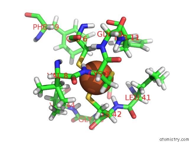
Mono view
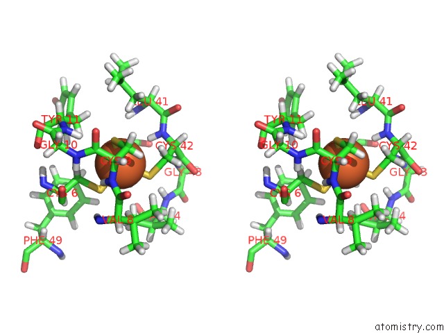
Stereo pair view
Mono view
Stereo pair view
A full contact list of Iron with other atoms in the Fe binding
site number 1 of Crystallographic Refinement of Rubredoxin at 1.2 Angstroms Resolution within 5.0Å range:
|
Reference:
K.D.Watenpaugh,
L.C.Sieker,
L.H.Jensen.
Crystallographic Refinement of Rubredoxin at 1 X 2 A Degrees Resolution. J.Mol.Biol. V. 138 615 1980.
ISSN: ISSN 0022-2836
PubMed: 7411618
DOI: 10.1016/S0022-2836(80)80020-9
Page generated: Mon Aug 5 09:33:29 2024
ISSN: ISSN 0022-2836
PubMed: 7411618
DOI: 10.1016/S0022-2836(80)80020-9
Last articles
Cl in 2XKDCl in 2XK7
Cl in 2XKC
Cl in 2XK8
Cl in 2XK4
Cl in 2XK6
Cl in 2XK3
Cl in 2XJZ
Cl in 2XJW
Cl in 2XJV