Iron »
PDB 4ryx-4toe »
4tlf »
Iron in PDB 4tlf: Crystal Structure of Thiol Dioxygenase From Pseudomonas Aeruginosa
Protein crystallography data
The structure of Crystal Structure of Thiol Dioxygenase From Pseudomonas Aeruginosa, PDB code: 4tlf
was solved by
M.Fellner,
E.P.Tchesnokov,
G.N.L.Jameson,
S.M.Wilbanks,
with X-Ray Crystallography technique. A brief refinement statistics is given in the table below:
| Resolution Low / High (Å) | 44.30 / 2.14 |
| Space group | P 41 21 2 |
| Cell size a, b, c (Å), α, β, γ (°) | 66.942, 66.942, 377.354, 90.00, 90.00, 90.00 |
| R / Rfree (%) | 19.8 / 25.1 |
Iron Binding Sites:
The binding sites of Iron atom in the Crystal Structure of Thiol Dioxygenase From Pseudomonas Aeruginosa
(pdb code 4tlf). This binding sites where shown within
5.0 Angstroms radius around Iron atom.
In total 4 binding sites of Iron where determined in the Crystal Structure of Thiol Dioxygenase From Pseudomonas Aeruginosa, PDB code: 4tlf:
Jump to Iron binding site number: 1; 2; 3; 4;
In total 4 binding sites of Iron where determined in the Crystal Structure of Thiol Dioxygenase From Pseudomonas Aeruginosa, PDB code: 4tlf:
Jump to Iron binding site number: 1; 2; 3; 4;
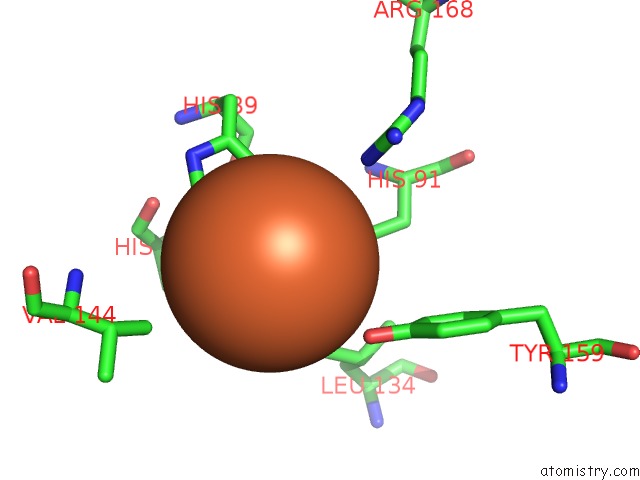
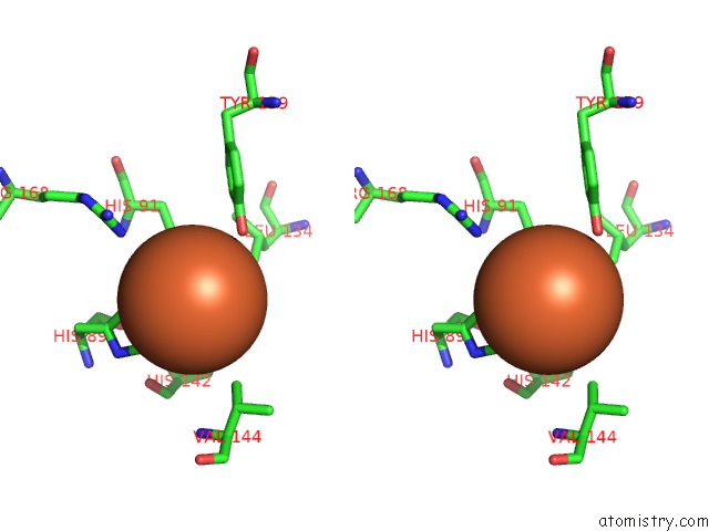
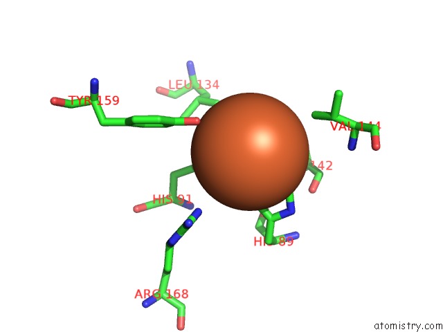
Iron binding site 1 out of 4 in 4tlf
Go back to
Iron binding site 1 out
of 4 in the Crystal Structure of Thiol Dioxygenase From Pseudomonas Aeruginosa

Mono view

Stereo pair view
Mono view
Stereo pair view
A full contact list of Iron with other atoms in the Fe binding
site number 1 of Crystal Structure of Thiol Dioxygenase From Pseudomonas Aeruginosa within 5.0Å range:
|
Iron binding site 2 out of 4 in 4tlf
Go back to
Iron binding site 2 out
of 4 in the Crystal Structure of Thiol Dioxygenase From Pseudomonas Aeruginosa
Mono view
Stereo pair view
Mono view
Stereo pair view
A full contact list of Iron with other atoms in the Fe binding
site number 2 of Crystal Structure of Thiol Dioxygenase From Pseudomonas Aeruginosa within 5.0Å range:
|
Iron binding site 3 out of 4 in 4tlf
Go back to
Iron binding site 3 out
of 4 in the Crystal Structure of Thiol Dioxygenase From Pseudomonas Aeruginosa

Mono view

Stereo pair view
Mono view
Stereo pair view
A full contact list of Iron with other atoms in the Fe binding
site number 3 of Crystal Structure of Thiol Dioxygenase From Pseudomonas Aeruginosa within 5.0Å range:
|
Iron binding site 4 out of 4 in 4tlf
Go back to
Iron binding site 4 out
of 4 in the Crystal Structure of Thiol Dioxygenase From Pseudomonas Aeruginosa
Mono view

Stereo pair view
Mono view
Stereo pair view
A full contact list of Iron with other atoms in the Fe binding
site number 4 of Crystal Structure of Thiol Dioxygenase From Pseudomonas Aeruginosa within 5.0Å range:
|
Reference:
E.P.Tchesnokov,
M.Fellner,
E.Siakkou,
T.Kleffmann,
L.W.Martin,
S.Aloi,
I.L.Lamont,
S.M.Wilbanks,
G.N.Jameson.
The Cysteine Dioxygenase Homologue From Pseudomonas Aeruginosa Is A 3-Mercaptopropionate Dioxygenase. J.Biol.Chem. V. 290 24424 2015.
ISSN: ESSN 1083-351X
PubMed: 26272617
DOI: 10.1074/JBC.M114.635672
Page generated: Mon Aug 5 10:20:38 2024
ISSN: ESSN 1083-351X
PubMed: 26272617
DOI: 10.1074/JBC.M114.635672
Last articles
Fe in 2YXOFe in 2YRS
Fe in 2YXC
Fe in 2YNM
Fe in 2YVJ
Fe in 2YP1
Fe in 2YU2
Fe in 2YU1
Fe in 2YQB
Fe in 2YOO