Iron »
PDB 4ryx-4toe »
4tnk »
Iron in PDB 4tnk: Rt Xfel Structure of Photosystem II 250 Microsec After the Third Illumination at 5.2 A Resolution
Enzymatic activity of Rt Xfel Structure of Photosystem II 250 Microsec After the Third Illumination at 5.2 A Resolution
All present enzymatic activity of Rt Xfel Structure of Photosystem II 250 Microsec After the Third Illumination at 5.2 A Resolution:
1.10.3.9;
1.10.3.9;
Protein crystallography data
The structure of Rt Xfel Structure of Photosystem II 250 Microsec After the Third Illumination at 5.2 A Resolution, PDB code: 4tnk
was solved by
J.Kern,
R.Tran,
R.Alonso-Mori,
S.Koroidov,
N.Echols,
J.Hattne,
M.Ibrahim,
S.Gul,
H.Laksmono,
R.G.Sierra,
R.J.Gildea,
G.Han,
J.Hellmich,
B.Lassalle-Kaiser,
R.Chatterjee,
A.Brewster,
C.A.Stan,
C.Gloeckner,
A.Lampe,
D.Difiore,
D.Milathianaki,
A.R.Fry,
M.M.Seibert,
J.E.Koglin,
E.Gallo,
J.Uhlig,
D.Sokaras,
T.-C.Weng,
P.H.Zwart,
D.E.Skinner,
M.J.Bogan,
M.Messerschmidt,
P.Glatzel,
G.J.Williams,
S.Boutet,
P.D.Adams,
A.Zouni,
J.Messinger,
N.K.Sauter,
U.Bergmann,
J.Yano,
V.K.Yachandra,
with X-Ray Crystallography technique. A brief refinement statistics is given in the table below:
| Resolution Low / High (Å) | 68.41 / 5.20 |
| Space group | P 21 21 21 |
| Cell size a, b, c (Å), α, β, γ (°) | 132.615, 229.296, 306.825, 90.00, 90.00, 90.00 |
| R / Rfree (%) | 27.1 / 28.9 |
Other elements in 4tnk:
The structure of Rt Xfel Structure of Photosystem II 250 Microsec After the Third Illumination at 5.2 A Resolution also contains other interesting chemical elements:
| Magnesium | (Mg) | 70 atoms |
| Manganese | (Mn) | 8 atoms |
| Chlorine | (Cl) | 2 atoms |
| Calcium | (Ca) | 6 atoms |
Iron Binding Sites:
The binding sites of Iron atom in the Rt Xfel Structure of Photosystem II 250 Microsec After the Third Illumination at 5.2 A Resolution
(pdb code 4tnk). This binding sites where shown within
5.0 Angstroms radius around Iron atom.
In total 6 binding sites of Iron where determined in the Rt Xfel Structure of Photosystem II 250 Microsec After the Third Illumination at 5.2 A Resolution, PDB code: 4tnk:
Jump to Iron binding site number: 1; 2; 3; 4; 5; 6;
In total 6 binding sites of Iron where determined in the Rt Xfel Structure of Photosystem II 250 Microsec After the Third Illumination at 5.2 A Resolution, PDB code: 4tnk:
Jump to Iron binding site number: 1; 2; 3; 4; 5; 6;
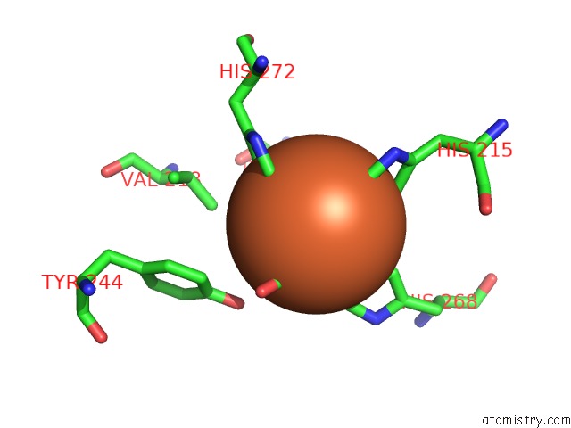
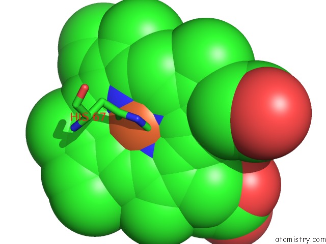
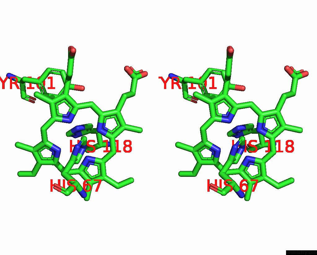
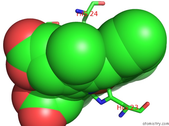
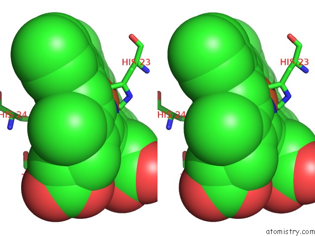
Iron binding site 1 out of 6 in 4tnk
Go back to
Iron binding site 1 out
of 6 in the Rt Xfel Structure of Photosystem II 250 Microsec After the Third Illumination at 5.2 A Resolution
Mono view
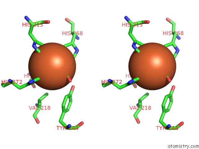
Stereo pair view
Mono view
Stereo pair view
A full contact list of Iron with other atoms in the Fe binding
site number 1 of Rt Xfel Structure of Photosystem II 250 Microsec After the Third Illumination at 5.2 A Resolution within 5.0Å range:
|
Iron binding site 2 out of 6 in 4tnk
Go back to
Iron binding site 2 out
of 6 in the Rt Xfel Structure of Photosystem II 250 Microsec After the Third Illumination at 5.2 A Resolution
Mono view
Stereo pair view
Mono view
Stereo pair view
A full contact list of Iron with other atoms in the Fe binding
site number 2 of Rt Xfel Structure of Photosystem II 250 Microsec After the Third Illumination at 5.2 A Resolution within 5.0Å range:
|
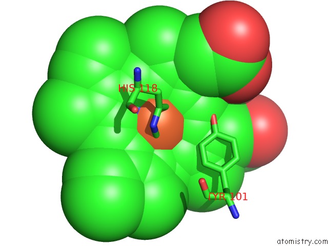
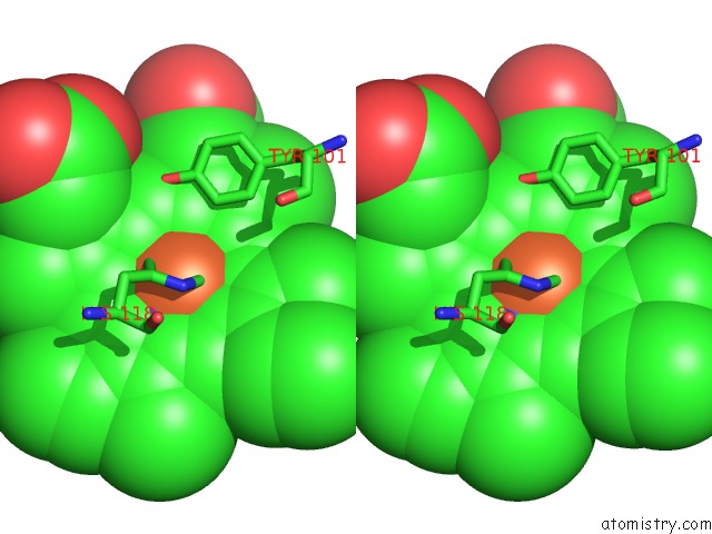
Iron binding site 3 out of 6 in 4tnk
Go back to
Iron binding site 3 out
of 6 in the Rt Xfel Structure of Photosystem II 250 Microsec After the Third Illumination at 5.2 A Resolution
Mono view
Stereo pair view
Mono view
Stereo pair view
A full contact list of Iron with other atoms in the Fe binding
site number 3 of Rt Xfel Structure of Photosystem II 250 Microsec After the Third Illumination at 5.2 A Resolution within 5.0Å range:
|
Iron binding site 4 out of 6 in 4tnk
Go back to
Iron binding site 4 out
of 6 in the Rt Xfel Structure of Photosystem II 250 Microsec After the Third Illumination at 5.2 A Resolution
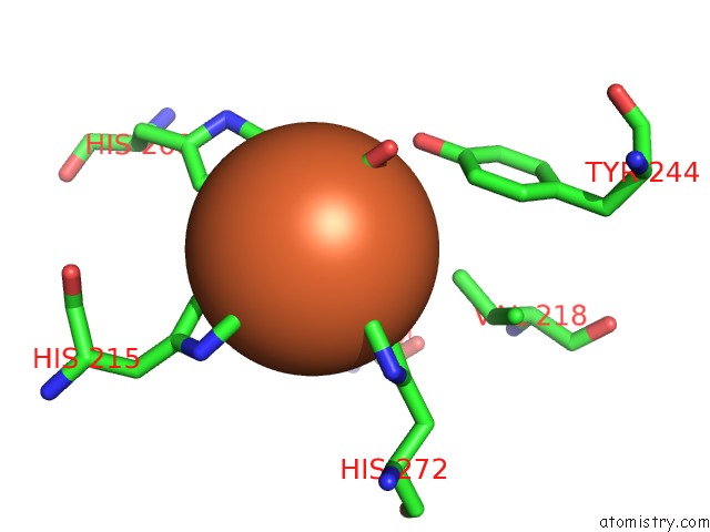
Mono view
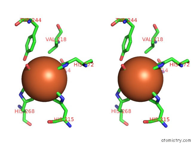
Stereo pair view
Mono view
Stereo pair view
A full contact list of Iron with other atoms in the Fe binding
site number 4 of Rt Xfel Structure of Photosystem II 250 Microsec After the Third Illumination at 5.2 A Resolution within 5.0Å range:
|
Iron binding site 5 out of 6 in 4tnk
Go back to
Iron binding site 5 out
of 6 in the Rt Xfel Structure of Photosystem II 250 Microsec After the Third Illumination at 5.2 A Resolution
Mono view
Stereo pair view
Mono view
Stereo pair view
A full contact list of Iron with other atoms in the Fe binding
site number 5 of Rt Xfel Structure of Photosystem II 250 Microsec After the Third Illumination at 5.2 A Resolution within 5.0Å range:
|
Iron binding site 6 out of 6 in 4tnk
Go back to
Iron binding site 6 out
of 6 in the Rt Xfel Structure of Photosystem II 250 Microsec After the Third Illumination at 5.2 A Resolution
Mono view
Stereo pair view
Mono view
Stereo pair view
A full contact list of Iron with other atoms in the Fe binding
site number 6 of Rt Xfel Structure of Photosystem II 250 Microsec After the Third Illumination at 5.2 A Resolution within 5.0Å range:
|
Reference:
J.Kern,
R.Tran,
R.Alonso-Mori,
S.Koroidov,
N.Echols,
J.Hattne,
M.Ibrahim,
S.Gul,
H.Laksmono,
R.G.Sierra,
R.J.Gildea,
G.Han,
J.Hellmich,
B.Lassalle-Kaiser,
R.Chatterjee,
A.S.Brewster,
C.A.Stan,
C.Glockner,
A.Lampe,
D.Difiore,
D.Milathianaki,
A.R.Fry,
M.M.Seibert,
J.E.Koglin,
E.Gallo,
J.Uhlig,
D.Sokaras,
T.C.Weng,
P.H.Zwart,
D.E.Skinner,
M.J.Bogan,
M.Messerschmidt,
P.Glatzel,
G.J.Williams,
S.Boutet,
P.D.Adams,
A.Zouni,
J.Messinger,
N.K.Sauter,
U.Bergmann,
J.Yano,
V.K.Yachandra.
Taking Snapshots of Photosynthetic Water Oxidation Using Femtosecond X-Ray Diffraction and Spectroscopy. Nat Commun V. 5 4371 2014.
ISSN: ESSN 2041-1723
PubMed: 25006873
DOI: 10.1038/NCOMMS5371
Page generated: Mon Aug 5 10:22:10 2024
ISSN: ESSN 2041-1723
PubMed: 25006873
DOI: 10.1038/NCOMMS5371
Last articles
Fe in 2YXOFe in 2YRS
Fe in 2YXC
Fe in 2YNM
Fe in 2YVJ
Fe in 2YP1
Fe in 2YU2
Fe in 2YU1
Fe in 2YQB
Fe in 2YOO