Iron »
PDB 4tof-4ud2 »
4u0p »
Iron in PDB 4u0p: The Crystal Structure of Lipoyl Synthase in Complex with S-Adenosyl Homocysteine
Enzymatic activity of The Crystal Structure of Lipoyl Synthase in Complex with S-Adenosyl Homocysteine
All present enzymatic activity of The Crystal Structure of Lipoyl Synthase in Complex with S-Adenosyl Homocysteine:
2.8.1.8;
2.8.1.8;
Protein crystallography data
The structure of The Crystal Structure of Lipoyl Synthase in Complex with S-Adenosyl Homocysteine, PDB code: 4u0p
was solved by
J.E.Harmer,
M.J.Hiscox,
J.Sandy,
P.C.Dinis,
P.L.Roach,
with X-Ray Crystallography technique. A brief refinement statistics is given in the table below:
| Resolution Low / High (Å) | 43.89 / 1.62 |
| Space group | C 2 2 21 |
| Cell size a, b, c (Å), α, β, γ (°) | 71.090, 161.130, 59.470, 90.00, 90.00, 90.00 |
| R / Rfree (%) | 17.8 / 20.7 |
Other elements in 4u0p:
The structure of The Crystal Structure of Lipoyl Synthase in Complex with S-Adenosyl Homocysteine also contains other interesting chemical elements:
| Sodium | (Na) | 1 atom |
Iron Binding Sites:
The binding sites of Iron atom in the The Crystal Structure of Lipoyl Synthase in Complex with S-Adenosyl Homocysteine
(pdb code 4u0p). This binding sites where shown within
5.0 Angstroms radius around Iron atom.
In total 8 binding sites of Iron where determined in the The Crystal Structure of Lipoyl Synthase in Complex with S-Adenosyl Homocysteine, PDB code: 4u0p:
Jump to Iron binding site number: 1; 2; 3; 4; 5; 6; 7; 8;
In total 8 binding sites of Iron where determined in the The Crystal Structure of Lipoyl Synthase in Complex with S-Adenosyl Homocysteine, PDB code: 4u0p:
Jump to Iron binding site number: 1; 2; 3; 4; 5; 6; 7; 8;
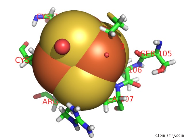
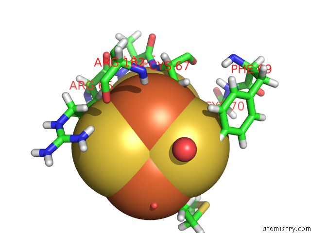
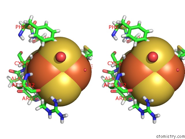
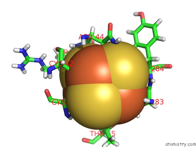
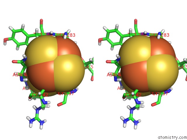
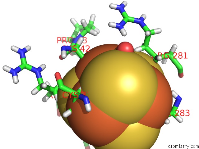
Iron binding site 1 out of 8 in 4u0p
Go back to
Iron binding site 1 out
of 8 in the The Crystal Structure of Lipoyl Synthase in Complex with S-Adenosyl Homocysteine
Mono view
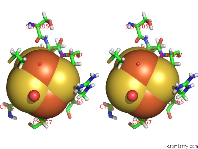
Stereo pair view
Mono view
Stereo pair view
A full contact list of Iron with other atoms in the Fe binding
site number 1 of The Crystal Structure of Lipoyl Synthase in Complex with S-Adenosyl Homocysteine within 5.0Å range:
|
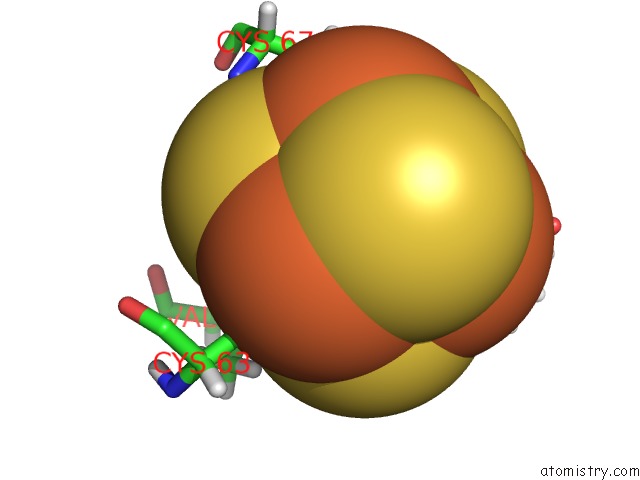
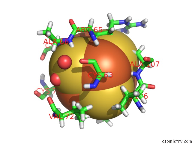
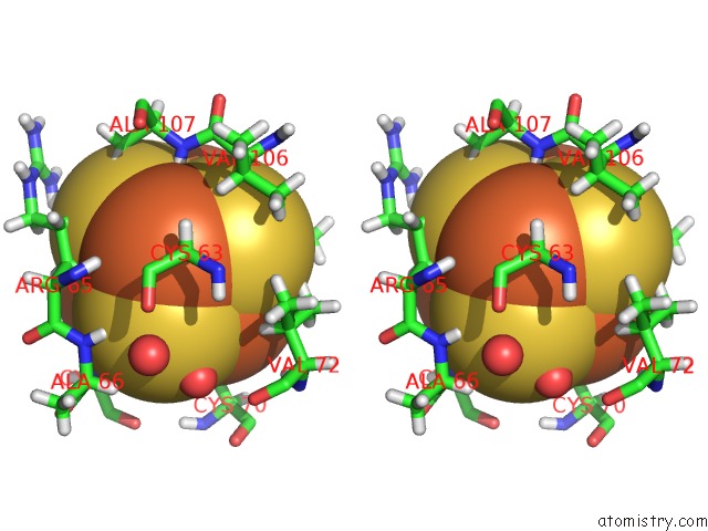
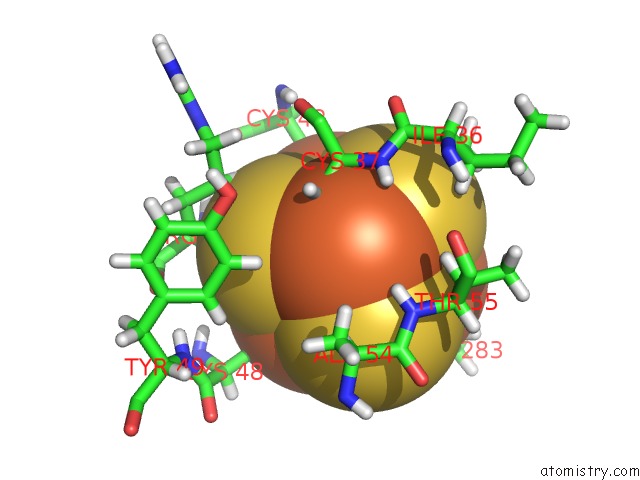
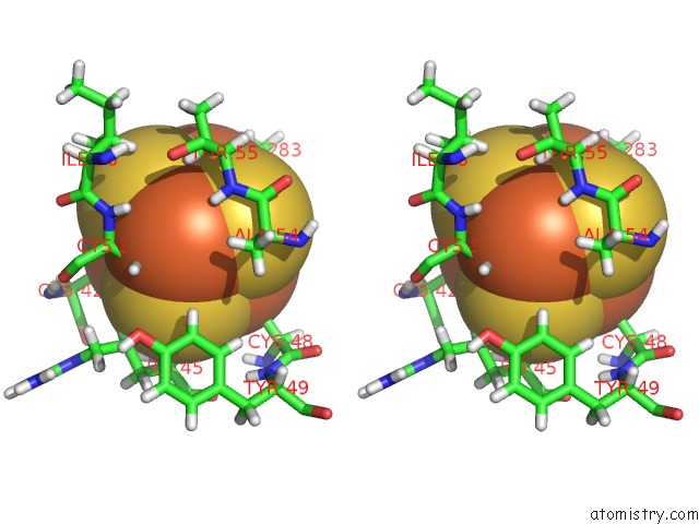
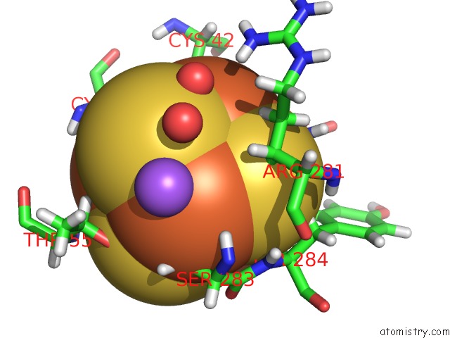
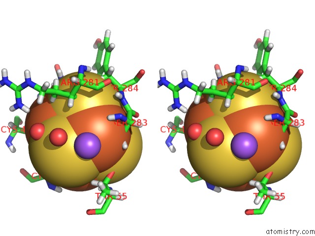
Iron binding site 2 out of 8 in 4u0p
Go back to
Iron binding site 2 out
of 8 in the The Crystal Structure of Lipoyl Synthase in Complex with S-Adenosyl Homocysteine
Mono view
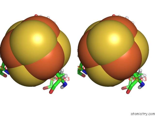
Stereo pair view
Mono view
Stereo pair view
A full contact list of Iron with other atoms in the Fe binding
site number 2 of The Crystal Structure of Lipoyl Synthase in Complex with S-Adenosyl Homocysteine within 5.0Å range:
|
Iron binding site 3 out of 8 in 4u0p
Go back to
Iron binding site 3 out
of 8 in the The Crystal Structure of Lipoyl Synthase in Complex with S-Adenosyl Homocysteine
Mono view
Stereo pair view
Mono view
Stereo pair view
A full contact list of Iron with other atoms in the Fe binding
site number 3 of The Crystal Structure of Lipoyl Synthase in Complex with S-Adenosyl Homocysteine within 5.0Å range:
|
Iron binding site 4 out of 8 in 4u0p
Go back to
Iron binding site 4 out
of 8 in the The Crystal Structure of Lipoyl Synthase in Complex with S-Adenosyl Homocysteine
Mono view
Stereo pair view
Mono view
Stereo pair view
A full contact list of Iron with other atoms in the Fe binding
site number 4 of The Crystal Structure of Lipoyl Synthase in Complex with S-Adenosyl Homocysteine within 5.0Å range:
|
Iron binding site 5 out of 8 in 4u0p
Go back to
Iron binding site 5 out
of 8 in the The Crystal Structure of Lipoyl Synthase in Complex with S-Adenosyl Homocysteine
Mono view
Stereo pair view
Mono view
Stereo pair view
A full contact list of Iron with other atoms in the Fe binding
site number 5 of The Crystal Structure of Lipoyl Synthase in Complex with S-Adenosyl Homocysteine within 5.0Å range:
|
Iron binding site 6 out of 8 in 4u0p
Go back to
Iron binding site 6 out
of 8 in the The Crystal Structure of Lipoyl Synthase in Complex with S-Adenosyl Homocysteine
Mono view
Stereo pair view
Mono view
Stereo pair view
A full contact list of Iron with other atoms in the Fe binding
site number 6 of The Crystal Structure of Lipoyl Synthase in Complex with S-Adenosyl Homocysteine within 5.0Å range:
|
Iron binding site 7 out of 8 in 4u0p
Go back to
Iron binding site 7 out
of 8 in the The Crystal Structure of Lipoyl Synthase in Complex with S-Adenosyl Homocysteine
Mono view

Stereo pair view
Mono view
Stereo pair view
A full contact list of Iron with other atoms in the Fe binding
site number 7 of The Crystal Structure of Lipoyl Synthase in Complex with S-Adenosyl Homocysteine within 5.0Å range:
|
Iron binding site 8 out of 8 in 4u0p
Go back to
Iron binding site 8 out
of 8 in the The Crystal Structure of Lipoyl Synthase in Complex with S-Adenosyl Homocysteine
Mono view
Stereo pair view
Mono view
Stereo pair view
A full contact list of Iron with other atoms in the Fe binding
site number 8 of The Crystal Structure of Lipoyl Synthase in Complex with S-Adenosyl Homocysteine within 5.0Å range:
|
Reference:
J.E.Harmer,
M.J.Hiscox,
P.C.Dinis,
S.J.Fox,
A.Iliopoulos,
J.E.Hussey,
J.Sandy,
F.T.Van Beek,
J.W.Essex,
P.L.Roach.
Structures of Lipoyl Synthase Reveal A Compact Active Site For Controlling Sequential Sulfur Insertion Reactions. Biochem.J. V. 464 123 2014.
ISSN: ESSN 1470-8728
PubMed: 25100160
DOI: 10.1042/BJ20140895
Page generated: Mon Aug 5 11:51:56 2024
ISSN: ESSN 1470-8728
PubMed: 25100160
DOI: 10.1042/BJ20140895
Last articles
Fe in 2BGVFe in 2BC5
Fe in 2BDM
Fe in 2BCN
Fe in 2B7S
Fe in 2B7R
Fe in 2B7H
Fe in 2B3Y
Fe in 2B24
Fe in 2B1X