Iron »
PDB 4tof-4ud2 »
4u99 »
Iron in PDB 4u99: Crystal Structure of An H-Nox Protein From S. Oneidensis in the Fe(II) Ligation State, Q154A/Q155A/K156A Mutant
Protein crystallography data
The structure of Crystal Structure of An H-Nox Protein From S. Oneidensis in the Fe(II) Ligation State, Q154A/Q155A/K156A Mutant, PDB code: 4u99
was solved by
M.A.Herzik Jr.,
R.Jonnalagadda,
J.Kuriyan,
M.A.Marletta,
with X-Ray Crystallography technique. A brief refinement statistics is given in the table below:
| Resolution Low / High (Å) | 47.88 / 2.00 |
| Space group | P 63 2 2 |
| Cell size a, b, c (Å), α, β, γ (°) | 164.003, 164.003, 101.718, 90.00, 90.00, 120.00 |
| R / Rfree (%) | 17.2 / 19.1 |
Other elements in 4u99:
The structure of Crystal Structure of An H-Nox Protein From S. Oneidensis in the Fe(II) Ligation State, Q154A/Q155A/K156A Mutant also contains other interesting chemical elements:
| Zinc | (Zn) | 2 atoms |
| Sodium | (Na) | 1 atom |
Iron Binding Sites:
The binding sites of Iron atom in the Crystal Structure of An H-Nox Protein From S. Oneidensis in the Fe(II) Ligation State, Q154A/Q155A/K156A Mutant
(pdb code 4u99). This binding sites where shown within
5.0 Angstroms radius around Iron atom.
In total 2 binding sites of Iron where determined in the Crystal Structure of An H-Nox Protein From S. Oneidensis in the Fe(II) Ligation State, Q154A/Q155A/K156A Mutant, PDB code: 4u99:
Jump to Iron binding site number: 1; 2;
In total 2 binding sites of Iron where determined in the Crystal Structure of An H-Nox Protein From S. Oneidensis in the Fe(II) Ligation State, Q154A/Q155A/K156A Mutant, PDB code: 4u99:
Jump to Iron binding site number: 1; 2;
Iron binding site 1 out of 2 in 4u99
Go back to
Iron binding site 1 out
of 2 in the Crystal Structure of An H-Nox Protein From S. Oneidensis in the Fe(II) Ligation State, Q154A/Q155A/K156A Mutant

Mono view
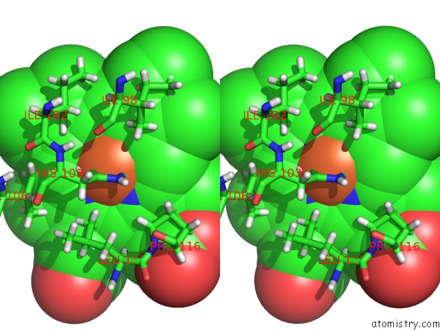
Stereo pair view
Mono view
Stereo pair view
A full contact list of Iron with other atoms in the Fe binding
site number 1 of Crystal Structure of An H-Nox Protein From S. Oneidensis in the Fe(II) Ligation State, Q154A/Q155A/K156A Mutant within 5.0Å range:
|
Iron binding site 2 out of 2 in 4u99
Go back to
Iron binding site 2 out
of 2 in the Crystal Structure of An H-Nox Protein From S. Oneidensis in the Fe(II) Ligation State, Q154A/Q155A/K156A Mutant

Mono view

Stereo pair view
Mono view
Stereo pair view
A full contact list of Iron with other atoms in the Fe binding
site number 2 of Crystal Structure of An H-Nox Protein From S. Oneidensis in the Fe(II) Ligation State, Q154A/Q155A/K156A Mutant within 5.0Å range:
|
Reference:
M.A.Herzik,
R.Jonnalagadda,
J.Kuriyan,
M.A.Marletta.
Structural Insights Into the Role of Iron-Histidine Bond Cleavage in Nitric Oxide-Induced Activation of H-Nox Gas Sensor Proteins. Proc.Natl.Acad.Sci.Usa V. 111 E4156 2014.
ISSN: ESSN 1091-6490
PubMed: 25253889
DOI: 10.1073/PNAS.1416936111
Page generated: Mon Aug 5 11:54:50 2024
ISSN: ESSN 1091-6490
PubMed: 25253889
DOI: 10.1073/PNAS.1416936111
Last articles
Zn in 9MJ5Zn in 9HNW
Zn in 9G0L
Zn in 9FNE
Zn in 9DZN
Zn in 9E0I
Zn in 9D32
Zn in 9DAK
Zn in 8ZXC
Zn in 8ZUF