Iron »
PDB 4tof-4ud2 »
4uax »
Iron in PDB 4uax: X-Ray Crystal Structure of Ligand Free CYP142A2 From Mycobacterium Smegmatis
Protein crystallography data
The structure of X-Ray Crystal Structure of Ligand Free CYP142A2 From Mycobacterium Smegmatis, PDB code: 4uax
was solved by
Y.Madrona,
with X-Ray Crystallography technique. A brief refinement statistics is given in the table below:
| Resolution Low / High (Å) | 19.59 / 1.78 |
| Space group | P 21 21 21 |
| Cell size a, b, c (Å), α, β, γ (°) | 56.650, 83.518, 94.501, 90.00, 90.00, 90.00 |
| R / Rfree (%) | 16.7 / 19.6 |
Iron Binding Sites:
The binding sites of Iron atom in the X-Ray Crystal Structure of Ligand Free CYP142A2 From Mycobacterium Smegmatis
(pdb code 4uax). This binding sites where shown within
5.0 Angstroms radius around Iron atom.
In total only one binding site of Iron was determined in the X-Ray Crystal Structure of Ligand Free CYP142A2 From Mycobacterium Smegmatis, PDB code: 4uax:
In total only one binding site of Iron was determined in the X-Ray Crystal Structure of Ligand Free CYP142A2 From Mycobacterium Smegmatis, PDB code: 4uax:
Iron binding site 1 out of 1 in 4uax
Go back to
Iron binding site 1 out
of 1 in the X-Ray Crystal Structure of Ligand Free CYP142A2 From Mycobacterium Smegmatis
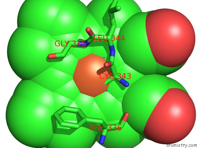
Mono view
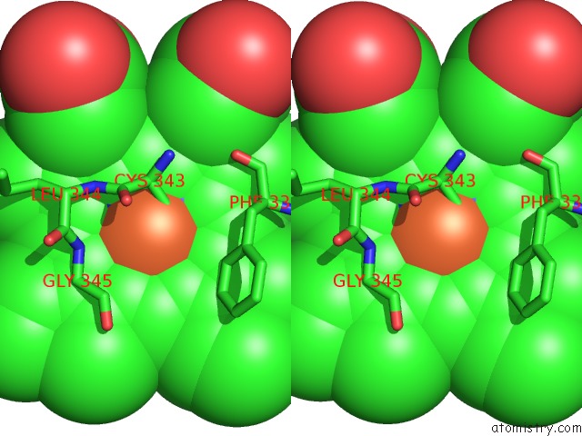
Stereo pair view
Mono view
Stereo pair view
A full contact list of Iron with other atoms in the Fe binding
site number 1 of X-Ray Crystal Structure of Ligand Free CYP142A2 From Mycobacterium Smegmatis within 5.0Å range:
|
Reference:
D.J.Frank,
Y.Madrona,
P.R.Ortiz De Montellano.
Cholesterol Ester Oxidation By Mycobacterial Cytochrome P450. J.Biol.Chem. V. 289 30417 2014.
ISSN: ESSN 1083-351X
PubMed: 25210044
DOI: 10.1074/JBC.M114.602771
Page generated: Mon Aug 5 12:00:25 2024
ISSN: ESSN 1083-351X
PubMed: 25210044
DOI: 10.1074/JBC.M114.602771
Last articles
Zn in 9J0NZn in 9J0O
Zn in 9J0P
Zn in 9FJX
Zn in 9EKB
Zn in 9C0F
Zn in 9CAH
Zn in 9CH0
Zn in 9CH3
Zn in 9CH1