Iron »
PDB 4ur2-4wgz »
4ur3 »
Iron in PDB 4ur3: Crystal Structure of the Pce Reductive Dehalogenase From S. Multivorans P2(1) Crystal Form
Enzymatic activity of Crystal Structure of the Pce Reductive Dehalogenase From S. Multivorans P2(1) Crystal Form
All present enzymatic activity of Crystal Structure of the Pce Reductive Dehalogenase From S. Multivorans P2(1) Crystal Form:
1.97.1.8;
1.97.1.8;
Protein crystallography data
The structure of Crystal Structure of the Pce Reductive Dehalogenase From S. Multivorans P2(1) Crystal Form, PDB code: 4ur3
was solved by
M.Bommer,
C.Kunze,
J.Fesseler,
T.Schubert,
G.Diekert,
H.Dobbek,
with X-Ray Crystallography technique. A brief refinement statistics is given in the table below:
| Resolution Low / High (Å) | 48.763 / 2.24 |
| Space group | P 1 21 1 |
| Cell size a, b, c (Å), α, β, γ (°) | 74.315, 110.601, 178.792, 90.00, 95.91, 90.00 |
| R / Rfree (%) | 17.93 / 21.77 |
Other elements in 4ur3:
The structure of Crystal Structure of the Pce Reductive Dehalogenase From S. Multivorans P2(1) Crystal Form also contains other interesting chemical elements:
| Cobalt | (Co) | 6 atoms |
Iron Binding Sites:
Pages:
>>> Page 1 <<< Page 2, Binding sites: 11 - 20; Page 3, Binding sites: 21 - 30; Page 4, Binding sites: 31 - 40; Page 5, Binding sites: 41 - 48;Binding sites:
The binding sites of Iron atom in the Crystal Structure of the Pce Reductive Dehalogenase From S. Multivorans P2(1) Crystal Form (pdb code 4ur3). This binding sites where shown within 5.0 Angstroms radius around Iron atom.In total 48 binding sites of Iron where determined in the Crystal Structure of the Pce Reductive Dehalogenase From S. Multivorans P2(1) Crystal Form, PDB code: 4ur3:
Jump to Iron binding site number: 1; 2; 3; 4; 5; 6; 7; 8; 9; 10;
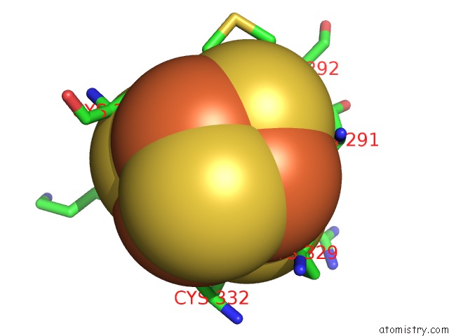
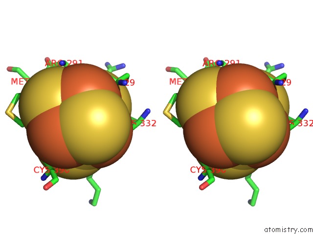
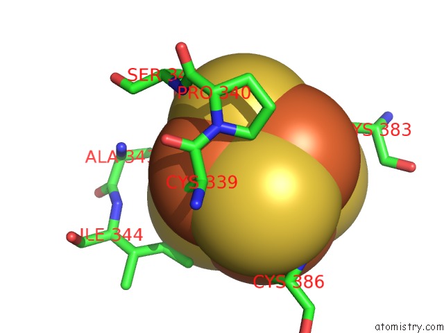
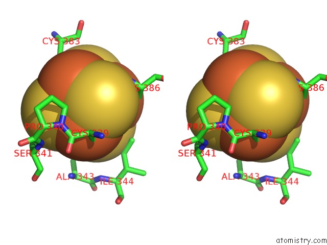
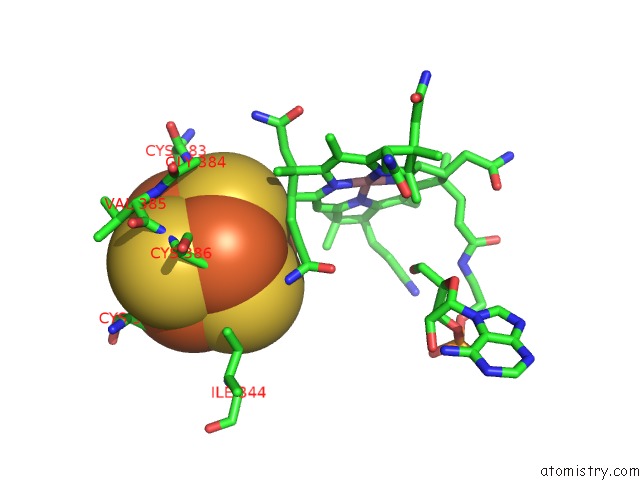
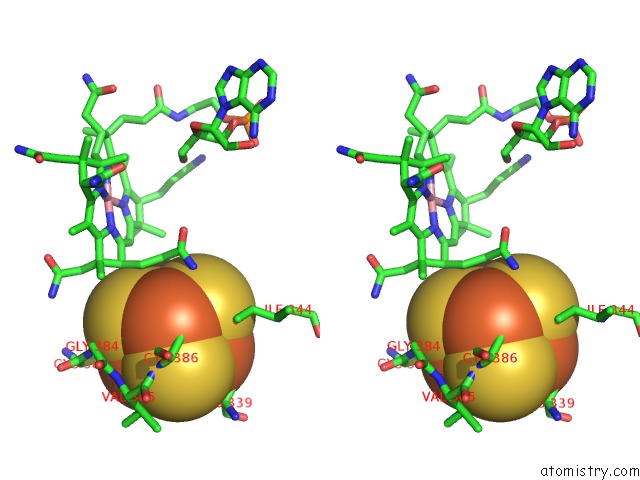
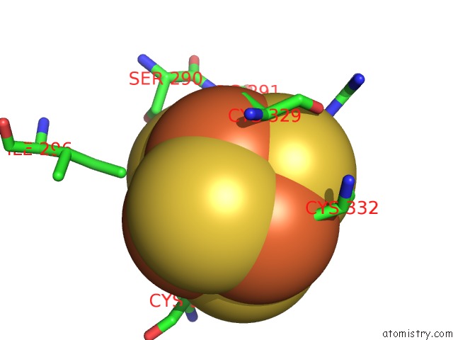
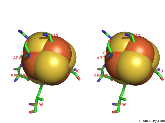
Iron binding site 1 out of 48 in 4ur3
Go back to
Iron binding site 1 out
of 48 in the Crystal Structure of the Pce Reductive Dehalogenase From S. Multivorans P2(1) Crystal Form
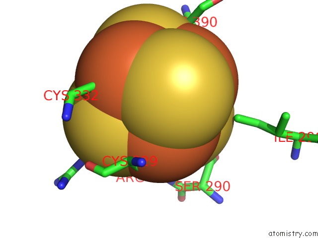
Mono view
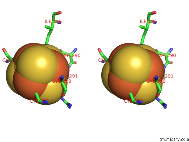
Stereo pair view
Mono view
Stereo pair view
A full contact list of Iron with other atoms in the Fe binding
site number 1 of Crystal Structure of the Pce Reductive Dehalogenase From S. Multivorans P2(1) Crystal Form within 5.0Å range:
|
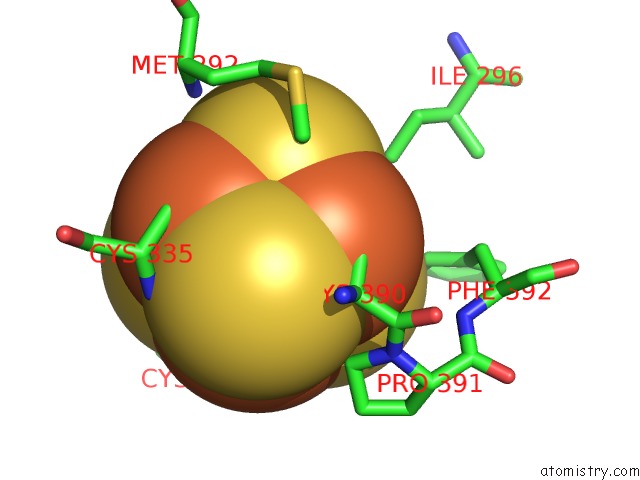
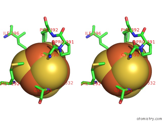
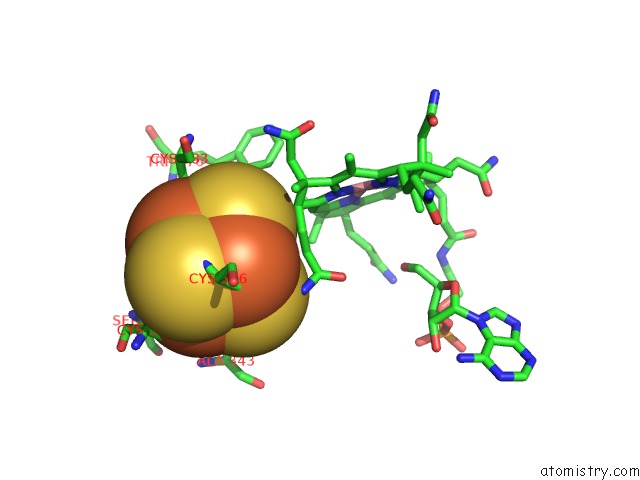
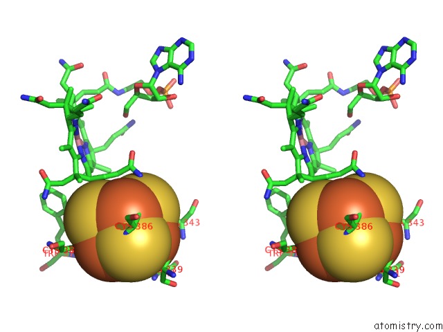
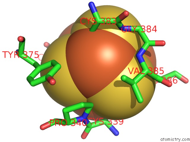
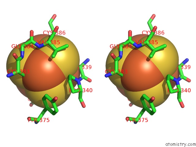
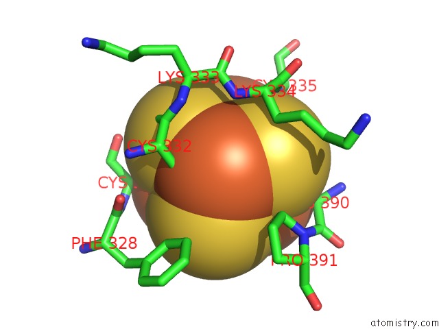
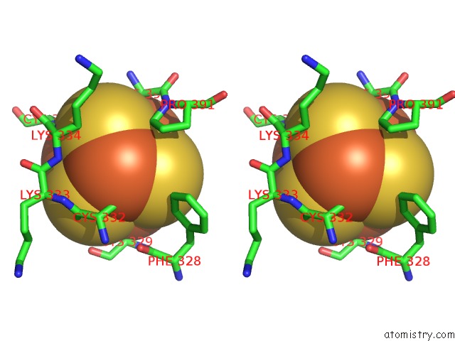
Iron binding site 2 out of 48 in 4ur3
Go back to
Iron binding site 2 out
of 48 in the Crystal Structure of the Pce Reductive Dehalogenase From S. Multivorans P2(1) Crystal Form
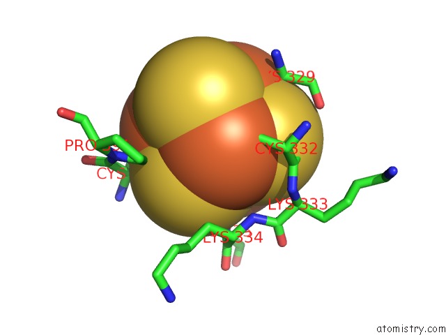
Mono view
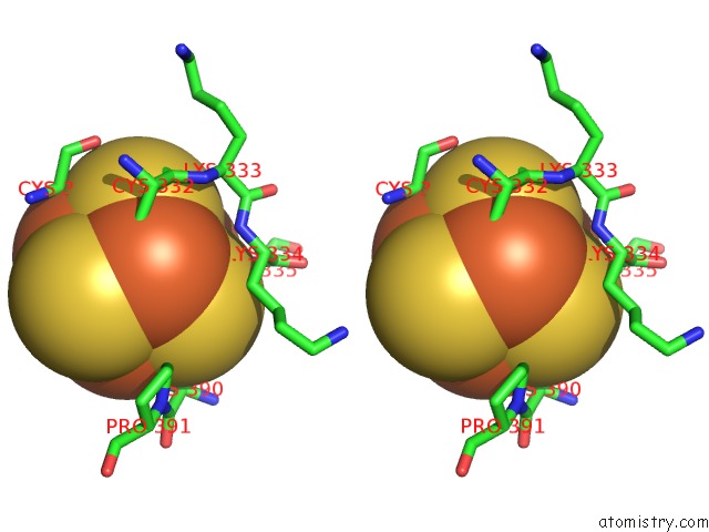
Stereo pair view
Mono view
Stereo pair view
A full contact list of Iron with other atoms in the Fe binding
site number 2 of Crystal Structure of the Pce Reductive Dehalogenase From S. Multivorans P2(1) Crystal Form within 5.0Å range:
|
Iron binding site 3 out of 48 in 4ur3
Go back to
Iron binding site 3 out
of 48 in the Crystal Structure of the Pce Reductive Dehalogenase From S. Multivorans P2(1) Crystal Form
Mono view
Stereo pair view
Mono view
Stereo pair view
A full contact list of Iron with other atoms in the Fe binding
site number 3 of Crystal Structure of the Pce Reductive Dehalogenase From S. Multivorans P2(1) Crystal Form within 5.0Å range:
|
Iron binding site 4 out of 48 in 4ur3
Go back to
Iron binding site 4 out
of 48 in the Crystal Structure of the Pce Reductive Dehalogenase From S. Multivorans P2(1) Crystal Form
Mono view
Stereo pair view
Mono view
Stereo pair view
A full contact list of Iron with other atoms in the Fe binding
site number 4 of Crystal Structure of the Pce Reductive Dehalogenase From S. Multivorans P2(1) Crystal Form within 5.0Å range:
|
Iron binding site 5 out of 48 in 4ur3
Go back to
Iron binding site 5 out
of 48 in the Crystal Structure of the Pce Reductive Dehalogenase From S. Multivorans P2(1) Crystal Form
Mono view
Stereo pair view
Mono view
Stereo pair view
A full contact list of Iron with other atoms in the Fe binding
site number 5 of Crystal Structure of the Pce Reductive Dehalogenase From S. Multivorans P2(1) Crystal Form within 5.0Å range:
|
Iron binding site 6 out of 48 in 4ur3
Go back to
Iron binding site 6 out
of 48 in the Crystal Structure of the Pce Reductive Dehalogenase From S. Multivorans P2(1) Crystal Form
Mono view
Stereo pair view
Mono view
Stereo pair view
A full contact list of Iron with other atoms in the Fe binding
site number 6 of Crystal Structure of the Pce Reductive Dehalogenase From S. Multivorans P2(1) Crystal Form within 5.0Å range:
|
Iron binding site 7 out of 48 in 4ur3
Go back to
Iron binding site 7 out
of 48 in the Crystal Structure of the Pce Reductive Dehalogenase From S. Multivorans P2(1) Crystal Form
Mono view
Stereo pair view
Mono view
Stereo pair view
A full contact list of Iron with other atoms in the Fe binding
site number 7 of Crystal Structure of the Pce Reductive Dehalogenase From S. Multivorans P2(1) Crystal Form within 5.0Å range:
|
Iron binding site 8 out of 48 in 4ur3
Go back to
Iron binding site 8 out
of 48 in the Crystal Structure of the Pce Reductive Dehalogenase From S. Multivorans P2(1) Crystal Form
Mono view
Stereo pair view
Mono view
Stereo pair view
A full contact list of Iron with other atoms in the Fe binding
site number 8 of Crystal Structure of the Pce Reductive Dehalogenase From S. Multivorans P2(1) Crystal Form within 5.0Å range:
|
Iron binding site 9 out of 48 in 4ur3
Go back to
Iron binding site 9 out
of 48 in the Crystal Structure of the Pce Reductive Dehalogenase From S. Multivorans P2(1) Crystal Form
Mono view
Stereo pair view
Mono view
Stereo pair view
A full contact list of Iron with other atoms in the Fe binding
site number 9 of Crystal Structure of the Pce Reductive Dehalogenase From S. Multivorans P2(1) Crystal Form within 5.0Å range:
|
Iron binding site 10 out of 48 in 4ur3
Go back to
Iron binding site 10 out
of 48 in the Crystal Structure of the Pce Reductive Dehalogenase From S. Multivorans P2(1) Crystal Form
Mono view
Stereo pair view
Mono view
Stereo pair view
A full contact list of Iron with other atoms in the Fe binding
site number 10 of Crystal Structure of the Pce Reductive Dehalogenase From S. Multivorans P2(1) Crystal Form within 5.0Å range:
|
Reference:
M.Bommer,
C.Kunze,
J.Fesseler,
T.Schubert,
G.Diekert,
H.Dobbek.
Structural Basis For Organohalide Respiration. Science V. 346 455 2014.
ISSN: ISSN 0036-8075
PubMed: 25278505
DOI: 10.1126/SCIENCE.1258118
Page generated: Mon Aug 5 14:05:31 2024
ISSN: ISSN 0036-8075
PubMed: 25278505
DOI: 10.1126/SCIENCE.1258118
Last articles
Cl in 3ZOQCl in 3ZNU
Cl in 3ZOA
Cl in 3ZO9
Cl in 3ZNX
Cl in 3ZNV
Cl in 3ZN5
Cl in 3ZNO
Cl in 3ZN6
Cl in 3ZMJ